37 uv vis block diagram
In mass spectrometry, matrix-assisted laser desorption/ionization (MALDI) is an ionization technique that uses a laser energy absorbing matrix to create ions from large molecules with minimal fragmentation. It has been applied to the analysis of biomolecules (biopolymers such as DNA, proteins, peptides and carbohydrates) and various organic molecules (such as polymers, … Install the Removable Terminal Block 12 Wire the Module 15 Communicate with the Module 17 Interpret Status Indicators 18 Specifications 20 . 2 POINT I/O 2 Current and 2 Voltage Input Analog Modules Publication 1734-IN027D-EN-E - December 2018 Important User Information Solid-state equipment has operational characteristics differing from those of electromechanical …
Block diagram of Heat Flux DSC. DSC is a commercially available instrument which has two (2) types: Heat Flux Type and Power Compensation Type. Figure 1 shows the block diagram of Heat Flux DSC as an example. Heat Flux DSC comprises the sample and reference holder, the heat resistor, the heat sink, and the heater. Heat of heater is supplied into the sample and the …
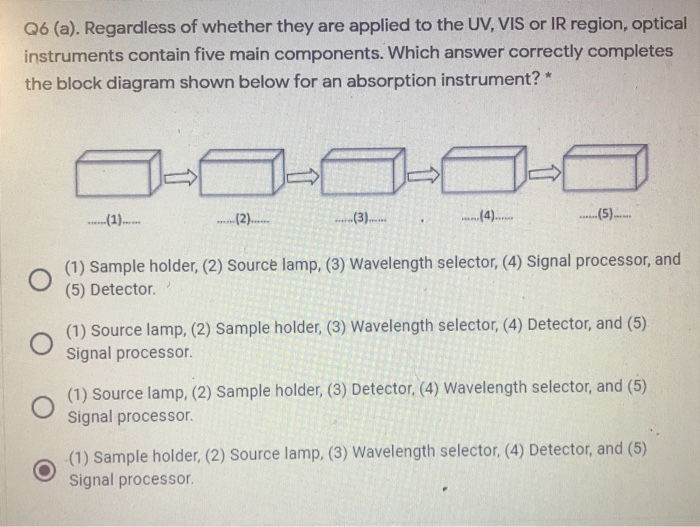
Uv vis block diagram
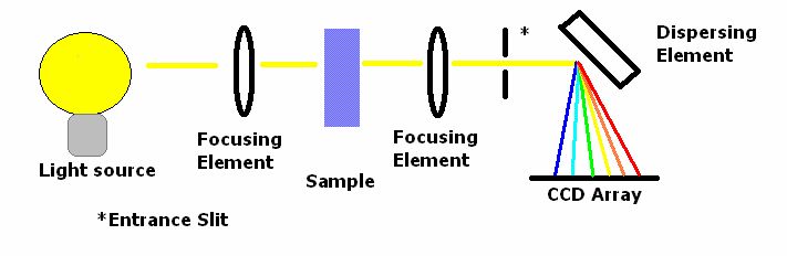
Diagram of a Czerny–Turner monochromator. A combined reflecting-focusing diffraction grating . A Fastie–Ebert monochromator. This is similar to the Czerny–Turner but uses a common collimator/refocusing mirror. In the common Czerny–Turner design, the broad-band illumination source (A) is aimed at an entrance slit (B). The amount of light energy available for use … The chart at the bottom displays the functional wavelength envelopes of the various detectors found in UV/Vis and UV/Vis/NIR spectrophotometers. Ultraviolet–visible spectroscopy - Wikipedia
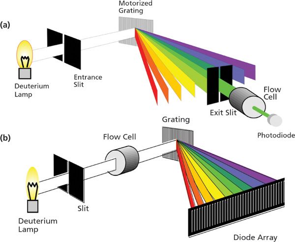
Uv vis block diagram. UV-VIS. BLUE-Wave – 200-1150nm; BLACK-Comet – concave grating; BLACK-Comet-SR – super range; SILVER-Nova – TE cooled sensitivity; Hi-Res – down to 0.1nm; HR-X eXtreme Hi-Res – 2x Resolutions – Hot for 2021! GREEN-Wave – .edu and teaching; NIR. DWARF-Star – 900-1700nm; RED-Wave-Micro – 1750-2150nm; High Res NIR – WDM to 0.4nm; RED-Wave … In Ultravoilet/Visible (UV-Vis) spectroscopy, electromagnetic radiation is emitted within the ... Schematic of our single beam diode array spectrometer. 12/08/2021 · As shown in the UV-Vis absorption spectra of the ILCP fabrics before and after UV irradiation (Supplementary Fig. 12), the peak intensity at 365 nm is corresponding to the π-π* transition of the ... 11 Sept 2021 — The simplest instrument for molecular UV/Vis absorption is a ... Schematic diagram of a fixed-wavelength, single-beam spectrophotometer.
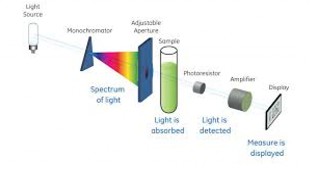
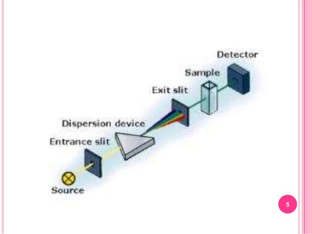
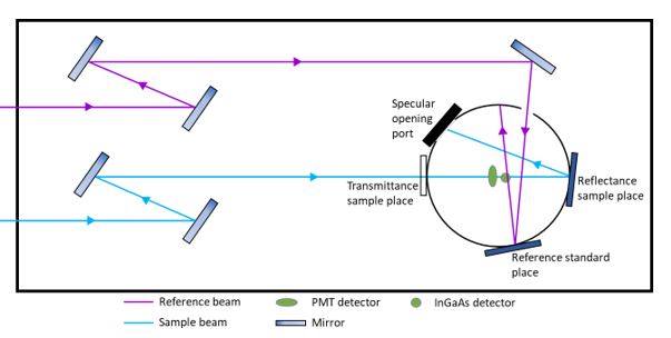
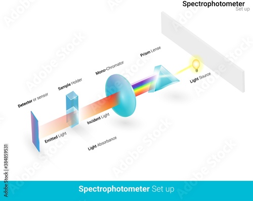
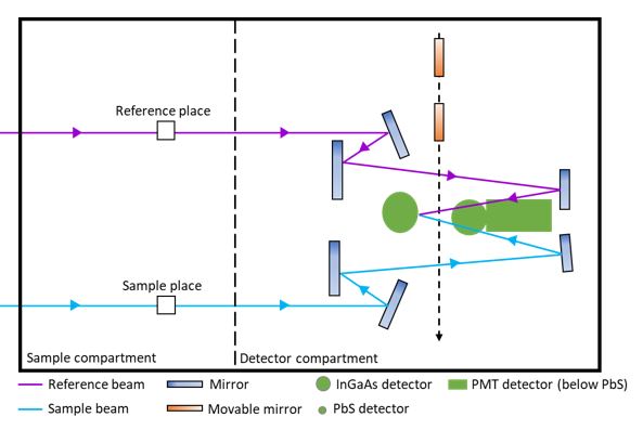
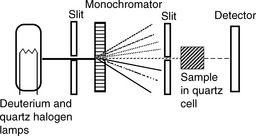
Download scientific diagram | Schematic diagram of UV-Visible Spectrophotometer from publication: A DETAILED STUDY ON OPTICAL AND PHYSICAL PROPERTIES OF ... In UV-Vis spectroscopy, wavelength is usually expressed in nanometers. (1 nm = 10-9 m). ... Schematic diagram of a double monochromator spectrophotometer.36 pages Block diagram of DTA. Figure 1 shows the block diagram of DTA. The sample and the reference are placed symmetrically in the furnace. The furnace is controlled under a temperature program and the temperature of the sample and the reference are changed. During this process, a differential thermocouple is set up to detect the temperature difference between the sample … A diagram of the components of a typical spectrometer are shown in the following diagram. The functioning of this instrument is relatively straightforward. A ...
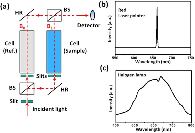
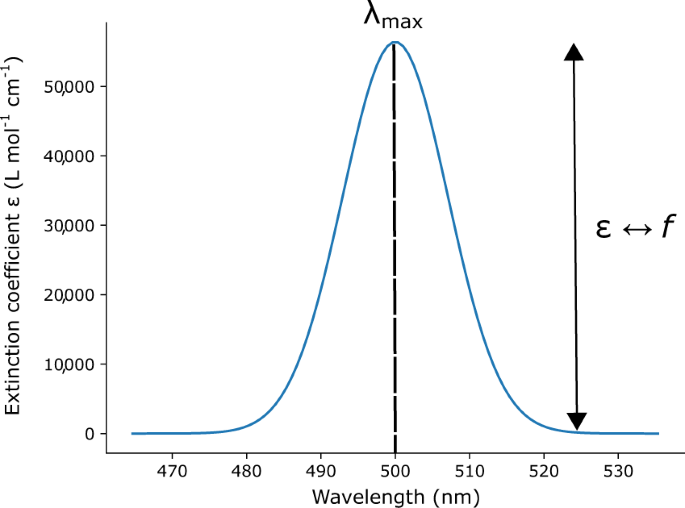
Have a look at this schematic diagram of a double-beam UV-Vis. spectrophotometer;. Instruments for measuring the absorption of U.V. or visible radiation are ... In a UV/Vis spectrum there is spectral resolution that is controlled by the instrument’s slit setting as well as data point resolution defined by the data point collection interval. First, we need to understand the characteristics of a well resolved absorbance peak generated by a typical spectrophotometer. At left we see a diagram of a single fully resolved absorbance peak. It … 20/04/2015 · circuit and an insect-attracting UV lamp of 365 nm wave-length. This ultraviolet This ultraviolet fluorescent lamp is m ounted in the middle of the cabine t … A point on the diagram represents a composition that is specified in terms of mole fraction or weight fraction. The point, (0.3, 0.4, 0.3) is at the center of the small triangle in the diagram and is located by following the red diagonal 60° line at red 0.3 and the horizontal line at blue 0.4 or any combination of two of the coordinates (A, B, C).
Ultraviolet–visible spectroscopy - Wikipedia

The chart at the bottom displays the functional wavelength envelopes of the various detectors found in UV/Vis and UV/Vis/NIR spectrophotometers.
Diagram of a Czerny–Turner monochromator. A combined reflecting-focusing diffraction grating . A Fastie–Ebert monochromator. This is similar to the Czerny–Turner but uses a common collimator/refocusing mirror. In the common Czerny–Turner design, the broad-band illumination source (A) is aimed at an entrance slit (B). The amount of light energy available for use …


0 Response to "37 uv vis block diagram"
Post a Comment