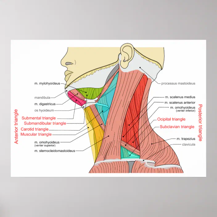
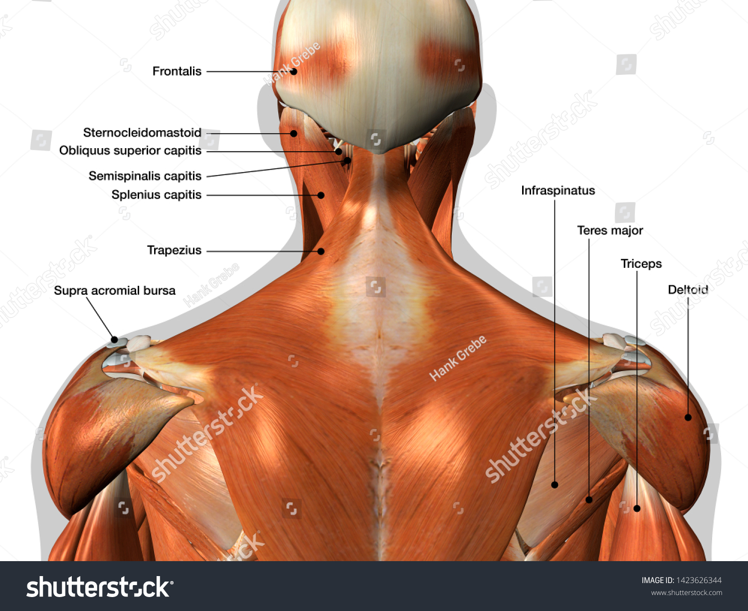
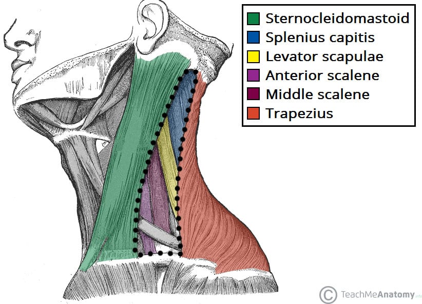
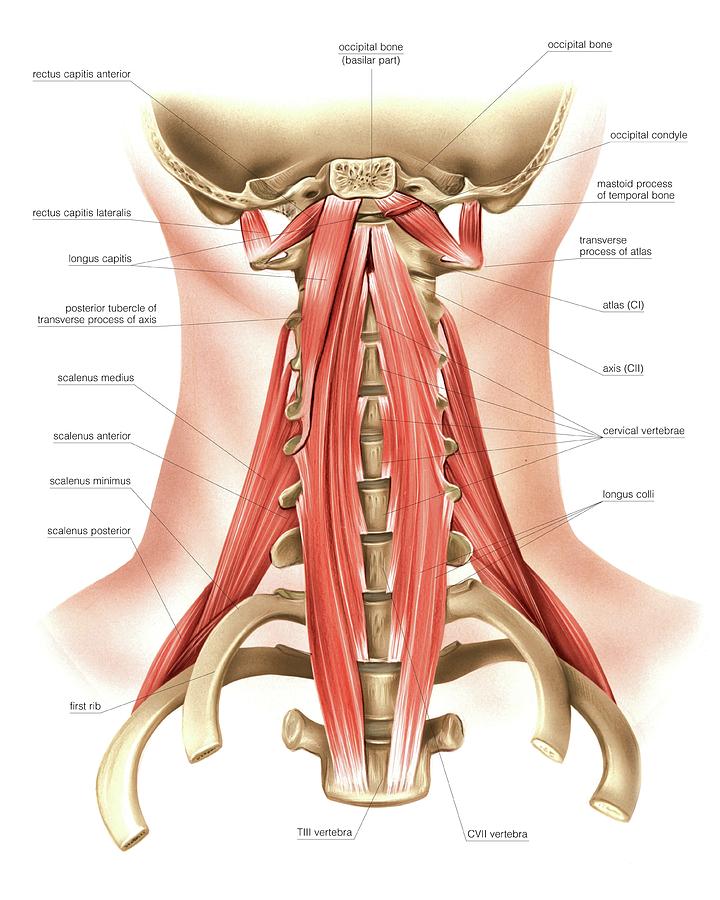
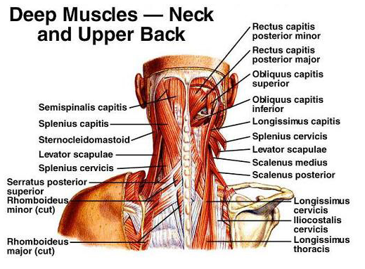
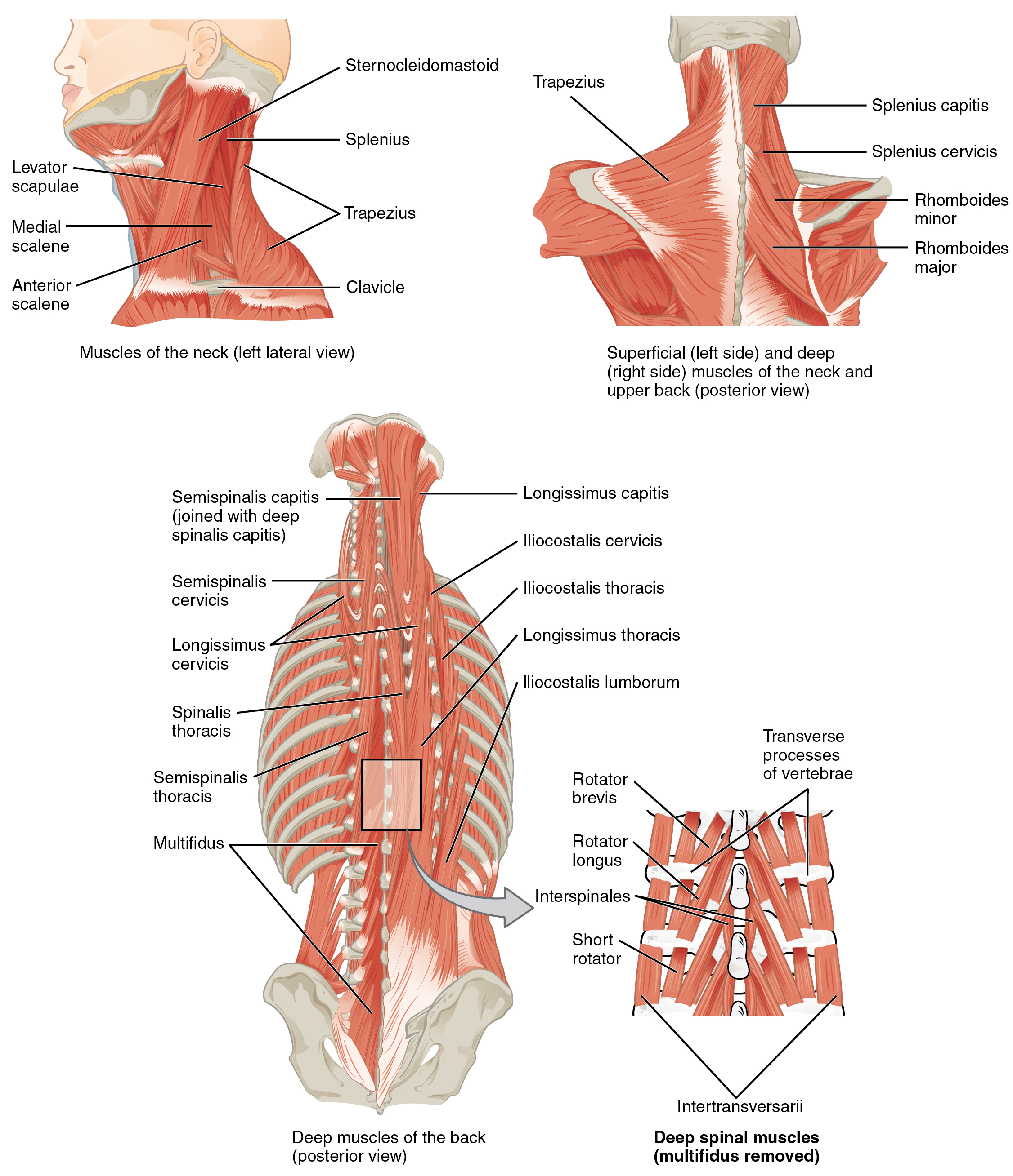
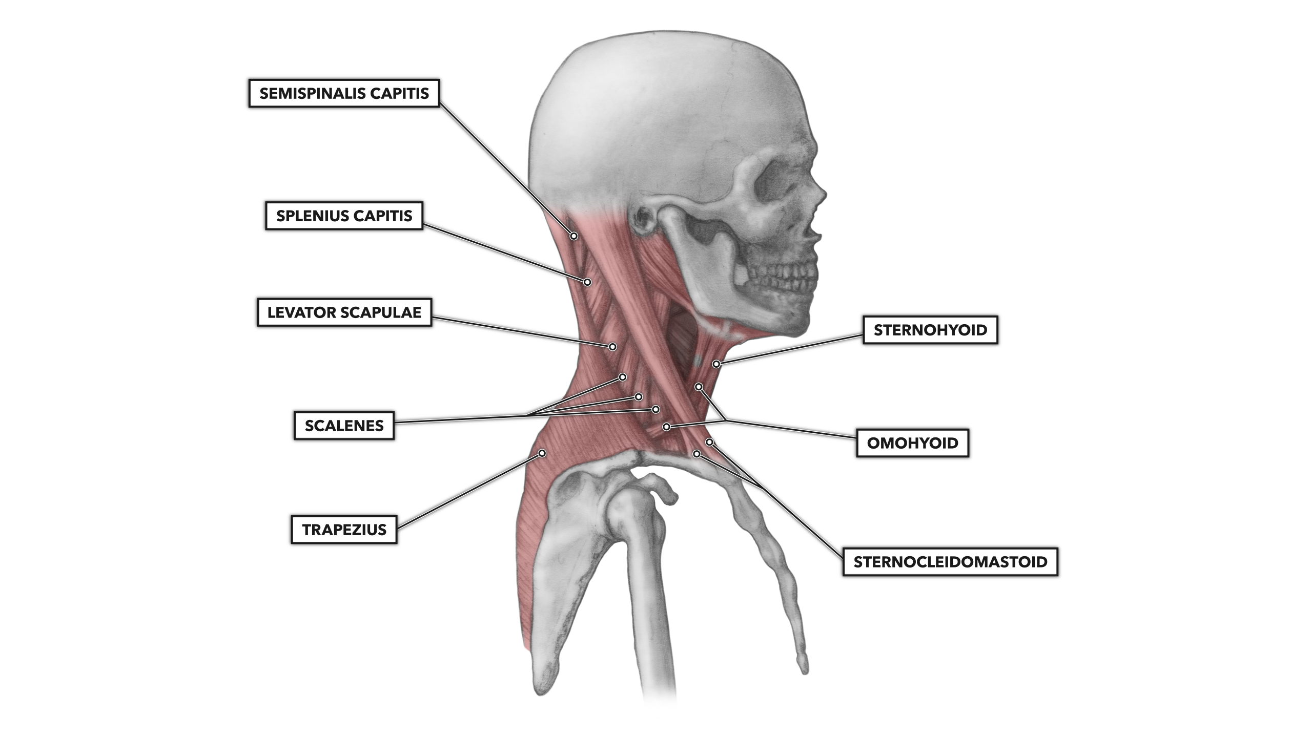
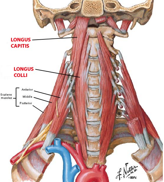
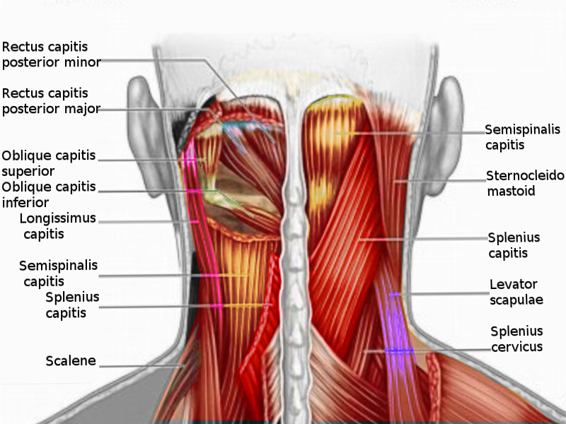
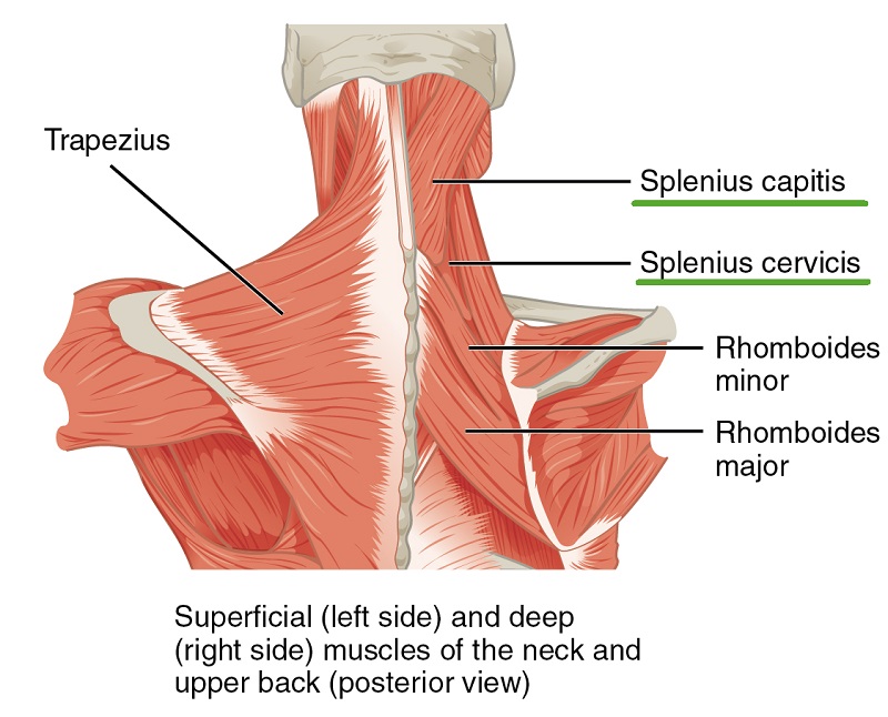
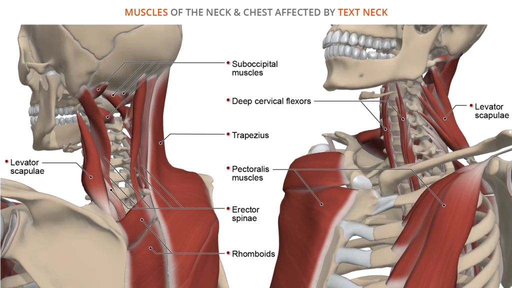
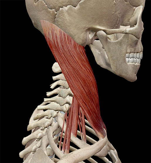
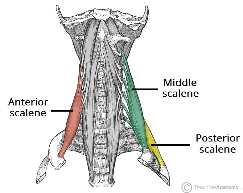
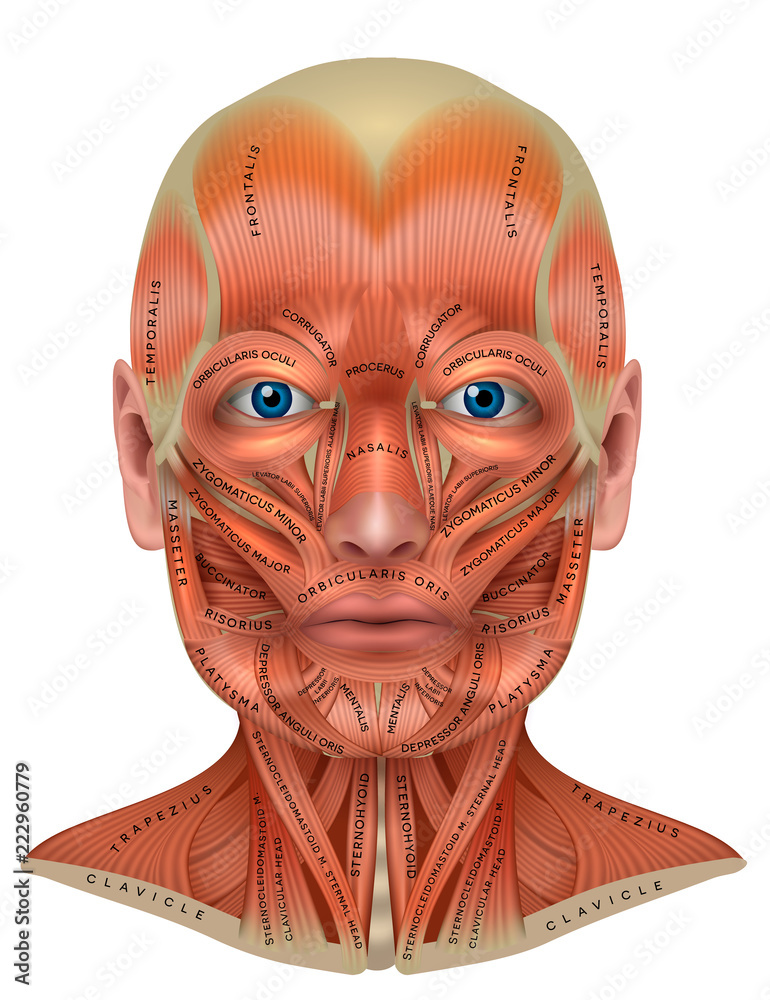
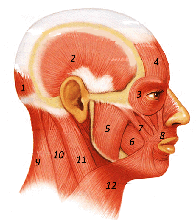
40 diagram of neck muscles
Anatomy of The Neck: Causes of Neck Pain and How to Manage ... Regardless of the cause, neck pain can interfere with daily life. Muscle Strain. When neck muscles are strained, whether from poor posture or an accident, the muscle stretches too far and can tear. This is also known as pulling a muscle. When a muscle is pulled more severely, inflammation can occur and swelling can develop. Nerve Compression Anatomy Of Neck And Shoulder Stock Photos, Pictures ... 3D illustration x-ray neck painful. Trapezius - Female Anatomy Muscles anatomy of neck and shoulder stock pictures, royalty-free photos & images. Trapezius - Female Anatomy Muscles. Human Arm and Torso of an Anatomical Model anatomy of neck and shoulder stock pictures, royalty-free photos & images.
Anatomy, Head and Neck, Cervical Nerves - NCBI Bookshelf Cervical nerves are spinal nerves that arise from the cervical region of the spinal cord. These nerves conduct motor and sensory information via efferent and afferent fibers, respectively, to and from the central nervous system. While classified as peripheral nerves, the motor cell body resides in the anterior horn of the spinal cord. There are eight pairs of cervical nerves, denoted C1 to C8 ...

Diagram of neck muscles
Neck muscles | Encyclopedia | Anatomy.app | Learn anatomy ... Neck muscles. The neck connects the head with the rest of the body, and it contains various tissue, blood vessels, nerves, lymphatics and organs, including many skeletal muscles. The neck muscles are responsible for movements of the neck and head. Also, these muscles provide structural support for the head. Muscle Charts of the Human Body - PT Direct Muscle Charts of the Human Body. For your reference value these charts show the major superficial and deep muscles of the human body. Neck Anatomy Pictures Bones, Muscles, Nerves May 06, 2015 · Here is a list of the many muscles that exist in the neck. Longus Colli & Capitis – Responsible for flexion of the head and neck. Rectus Capitis Anterior – Responsible for flexion of the neck Rectus Capitis Lateralis – Helps the neck to bend to the side. Scalene Muscles – Responsible for lifting the first and second ribs i.e. assist with breathing
Diagram of neck muscles. PDF 1 2 neck stretches - Boulder Therapeutics MUSCLES STRETCHED:This is a great warm-up stretch for all neck muscles. HINT: Throughout these routines, think about reach-ing your neck up and out before bending it to the sides, front or back. Elongate the stretched side with particular attention to avoid "crunch-ing" the shortened side. CHAPTER 3: NECK STRETCHES BOULDER THERAPEUTICS, INC. Muscles of the Neck - TeachMeAnatomy The muscles of the neck are present in four main groups. The suboccipital muscles act to rotate the head and extend the neck.Rectus capitis posterior major and Rectus capitis posterior minor attach the inferior nuchal line of the occiput to the C2 and C1 vertebrae respectively.Obliquus capitis superior also extends from the occiput to C1 while obliquus capitis inferior originates from C2 and ... Back Muscles: Names And Diagram - Science Trends The levatores costarum muscles are 12 small muscles which, from the upper 11th thoracic vertebrae and the seventh cervical vertebrae, pass downwards and run into the outer portion of the ribs below the vertebrae. They help the lungs move and assist in the process of breathing. The interspinales muscles are pairs of muscles located on both sides of the interspinal ligament. Neck muscle phenotypes in Tbx1 and Pax3 mutants. (A-I ... Download scientific diagram | Neck muscle phenotypes in Tbx1 and Pax3 mutants. (A-I) Immunostainings for Tnnt3 on coronal cryosections of control, Tbx1-null and Pax3-null fetuses at E18.5 (n = 3 ...
lateral neck muscles. | Download Scientific Diagram Download scientific diagram | lateral neck muscles. from publication: Quality of life in cervical dystonia after treatment with botulinum toxin A: a 24-week prospective study | Objective This ... Anatomy, Head and Neck, Mastication Muscles - StatPearls ... The primary muscles of mastication (chewing food) are the temporalis, medial pterygoid, lateral pterygoid, and masseter muscles. The four main muscles of mastication attach to the rami of the mandible and function to move the jaw (mandible). The cardinal mandibular movements of mastication are elevation, depression, protrusion, retraction, and side to side movement. Head and neck muscles labeled anatomical diagram, facial ... Head and neck muscles labeled anatomical diagram, facial vector illustration with female face, health care educational information poster € 7.99 Muscles of the Head and Neck - Anatomy Pictures and ... The neck muscles, including the sternocleidomastoid and the trapezius, are responsible for the gross motor movement in the muscular system of the head and neck. They move the head in every direction, pulling the skull and jaw towards the shoulders, spine, and scapula. Working in pairs on the left and right sides of the body, these muscles ...
Back Muscles: Anatomy of Upper, Middle & Lower Back Pain ... Sternocleidomastoids. The sternocleidomastoids are strong, large muscles located on either side of the neck. Individually, they rotate the head left or right. Together, they flex or bend the head towards the chest. These muscles start at the breastbone (sternum) and collarbone (clavicle) and end behind the ears (at the mastoid process of the temporal bone of the skull). Of The Best Anatomy Muscle Labeling Worksheet - The ... Math Coloring Worksheets 3rd Grade. 6 best images of printable worksheets muscle anatomy blank head and neck muscles diagram muscular system diagram worksheet and label muscles worksheet practice sheet for name the parts of the human brain bing images strength is the product of struggle you must do what others dont to acheive what others wont. Shoulder Pain Diagram: Diagnosis Chart - Shoulder Pain Exp A. Muscle Strain. Straining the upper trapezius muscles is a common cause of pain across the top part of the back of the shoulders. You may have overworked the muscles doing heavy lifting or playing racket sports or may store tension in your upper traps if you spend all day in-front of the computer. LEARN MORE > Neck Problems Chin & Neck Muscles Diagram | Quizlet Start studying Chin & Neck Muscles. Learn vocabulary, terms, and more with flashcards, games, and other study tools.
Neck Muscles Anatomy, Diagram & Pictures | Body Maps Jan 20, 2018 · Neck muscles are bodies of tissue that produce motion in the neck when stimulated. The muscles of the neck run from the base of the skull to the upper back and work together to bend the head and ...
Neck muscles anatomy: List, origins, insertions, action ... Muscles of the neck (Musculi cervicales) The muscles of the neck are muscles that cover the area of the neck.These muscles are mainly responsible for the movement of the head in all directions. They consist of 3 main groups of muscles: anterior, lateral and posterior groups, based on their position in the neck.The musculature of the neck is further divided into more specific groups based on a ...
Diy Skeletal Muscle Diagram Worksheet - Labelco Featuring 10 of the more widely-known muscles including abdominals pectorals and hamstrings this labelling activity makes a great addition to any. Human body muscle diagram worksheet label muscles worksheet and blank head and neck muscles diagram are three of main things we want to present to you based on the post title.
Instant Anatomy - Head and Neck - Muscles Instant anatomy is a specialised web site for you to learn all about human anatomy of the body with diagrams, podcasts and revision questions
PDF Anatomy & Physiology Cat Muscles Ventral view 1 Deep muscles of the thigh (lateral view), Tensor fascia Iata Biceps femoris Vastus lateral's . Sup raspinatus Rhomboideus Infraspinatus Teres major C/avotrape zius C/avobrachia/is deltoidius A trapezius Latissimus dorsi Tensor Facia Latae Bicepts Femoñs Gastmcnemius Calcaneal Tendon Semitendinosis .
Dog Neck Anatomy - Bones, Muscle, Glands, Veins, and Other ... The dog neck's most vital structures and organs are the superficial muscles, neck bones, thyroid glands, esophagus, trachea, blood vessels (artery and veins), and lymph nodes. So, my goal is to provide explicit knowledge on these structures and organs from the dog neck.
Neck Muscles Anatomy, Function & Diagram | Body Maps Feb 17, 2015 · Located underneath the platysma on the sides of the neck are the sternocleidomastoid muscles. With one on each side of the neck, these help flex the neck and rotate the head upward and side to...
Cow Anatomy - External Body Parts and Internal Organs with ... The most important muscles of the neck region of a cow - here, you will find brachiocephalic, omotransverasarius, sternocephalicus, cervical part of trapezius, rhomboideus, and more. Again, the most important muscles of the thoracic limb of a cow - brachicephalicus, omotransversarius, pectoral muscle, muscle of shoulder, muscles of arm, and ...
Neck and Shoulder Pain Anatomy - selfcare4rsi.com Neck and Shoulder Pain Anatomy. The anatomy of the neck and shoulders is very interesting. These critical parts of the upper body are very prone to developing pain because the position of all the bones in the neck and shoulders are completely dependent on the balance and alignment of the muscles and fascia that lash them together and allow for movement between them.
Neck Exercises: Dos and Don'ts - WebMD You can work your neck muscles like any other muscles. Stretches work, but you can also do simple exercises like the ones below. They can improve your neck strength and your range of motion.
Neck Anatomy: Muscles, glands, organs | Kenhub Neck spaces. The content of the neck is grouped into 4 neck spaces, called the compartments.. Vertebral compartment: contains cervical vertebrae and postural muscles.; Visceral compartment: contains glands (thyroid, parathyroid, and thymus), the larynx, pharynx and trachea.; Two vascular compartments: contain the common carotid artery, internal jugular vein and the vagus nerve, on each side of ...
Neck Anatomy Pictures Bones, Muscles, Nerves May 06, 2015 · Here is a list of the many muscles that exist in the neck. Longus Colli & Capitis – Responsible for flexion of the head and neck. Rectus Capitis Anterior – Responsible for flexion of the neck Rectus Capitis Lateralis – Helps the neck to bend to the side. Scalene Muscles – Responsible for lifting the first and second ribs i.e. assist with breathing
Muscle Charts of the Human Body - PT Direct Muscle Charts of the Human Body. For your reference value these charts show the major superficial and deep muscles of the human body.
Neck muscles | Encyclopedia | Anatomy.app | Learn anatomy ... Neck muscles. The neck connects the head with the rest of the body, and it contains various tissue, blood vessels, nerves, lymphatics and organs, including many skeletal muscles. The neck muscles are responsible for movements of the neck and head. Also, these muscles provide structural support for the head.

















:background_color(FFFFFF):format(jpeg)/images/library/12564/neck-viscera-cadaver.png)
0 Response to "40 diagram of neck muscles"
Post a Comment