38 atomic force microscope diagram
Atomic Force Microscope Market Analysis and Demand with Forecast Overview To 2028. The Atomic Force Microscope Market report is compilation of intelligent, broad research studies that will help players and stakeholders to make informed business decisions in future. It offers detailed research and analysis of key aspects of the Atomic Force ... Transmission electron microscopy (TEM) is a microscopy technique in which a beam of electrons is transmitted through a specimen to form an image. The specimen is most often an ultrathin section less than 100 nm thick or a suspension on a grid.
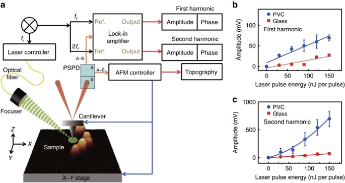
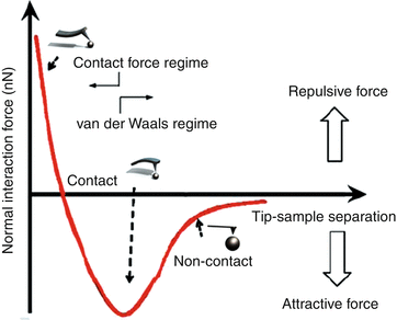
Atomic Force Microscopy (AFM) 1. General Principle The Atomic Force Microscope is a kind of scanning probe microscope in which a topographical image of the sample surface can be achieved based on the interactions between a tip and a sample surface. The atomic force microscope was invented by Gerd Binning et al. in 1986 at IBM Zurich based on ...

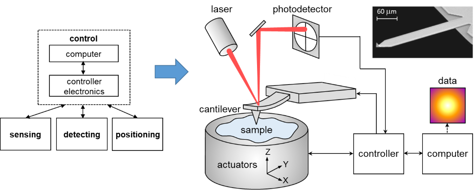
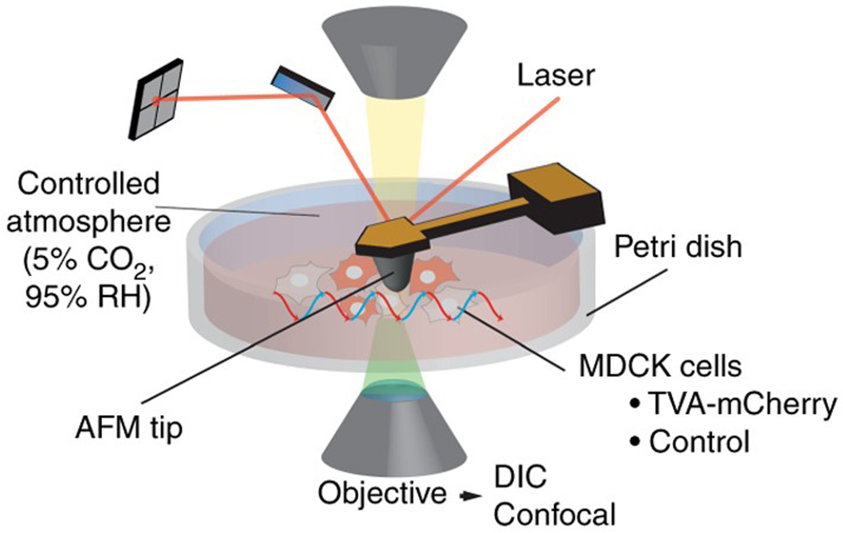
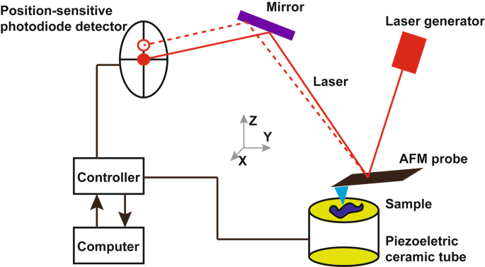
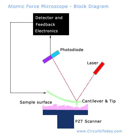
Atomic force microscope diagram
Visualization of solvation structure on Li 4 Ti 5 O 12 (111)/ ionic liquid-based electrolyte interface by atomic force microscopy Yifan Bao, Mitsunori Kitta, Takashi Ichii et al.-Solvation structure on water-in-salt/mica interfaces and its molality dependence investigated by atomic force microscopy Takashi Ichii, Satoshi Ichikawa, Yuya Yamada ... Functionalizing the tip of an atomic force microscope with a CO molecule enabled atomic-resolution imaging of single molecules, and measurement of their adsorption geometry and bond-order relations. In addition, by using scanning tunneling microscopy and Kelvin probe force microscopy, the density of the molecular frontier orbitals and the ... To learn more about Atomic Force Microscopy, click through. AFM microscopes are among the best solutions for measuring the nanoscale surface metrology and material properties of samples. A conventional compound light microscope is limited to a maximum sample magnification of approximately 1000x; a quantity that is dictated by the wavelengths of ...
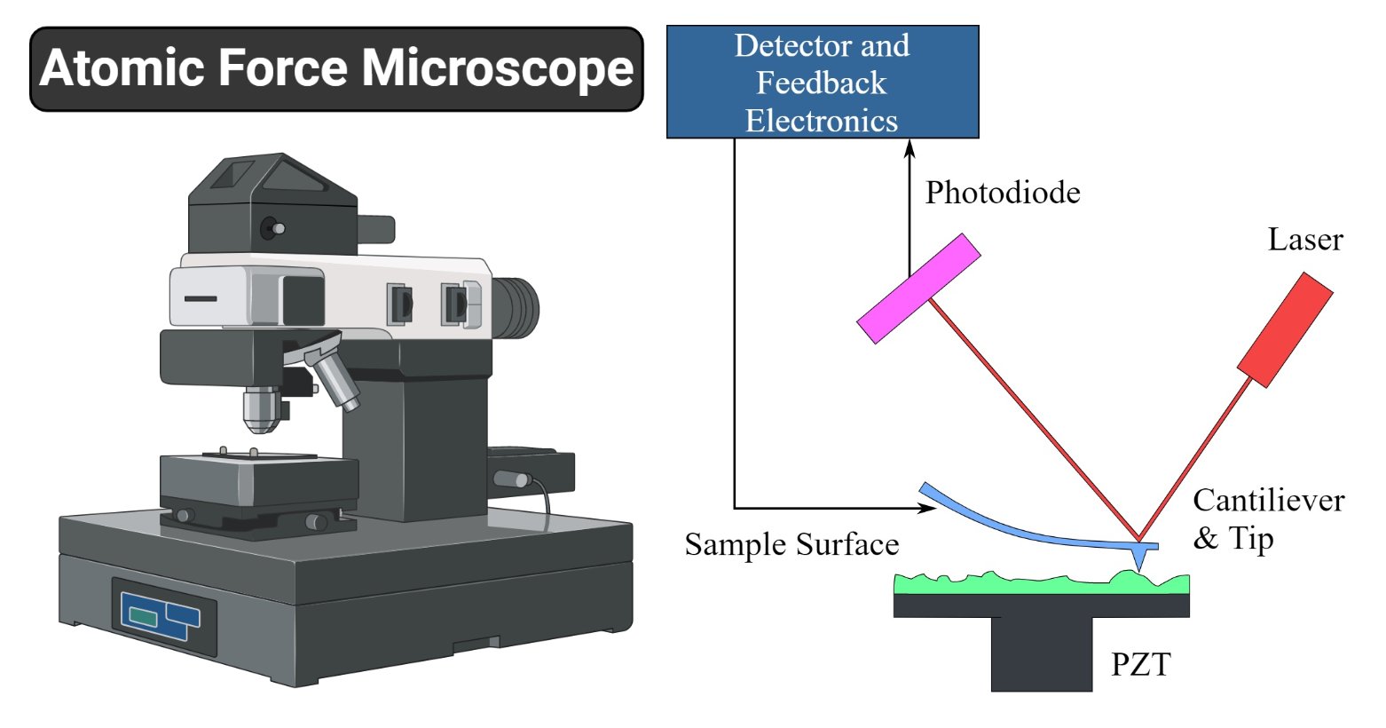
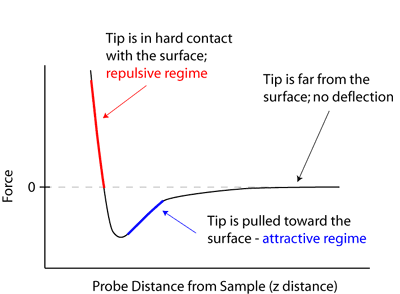
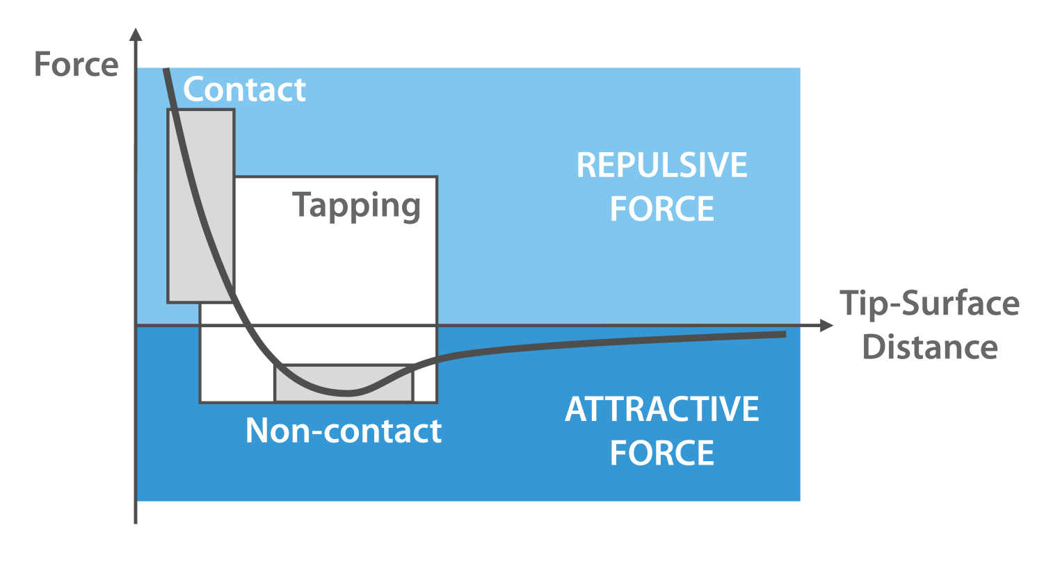
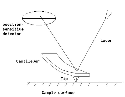
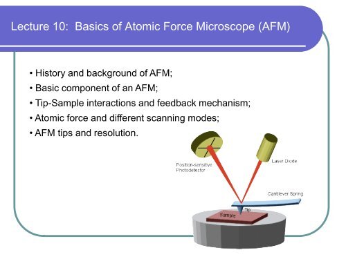
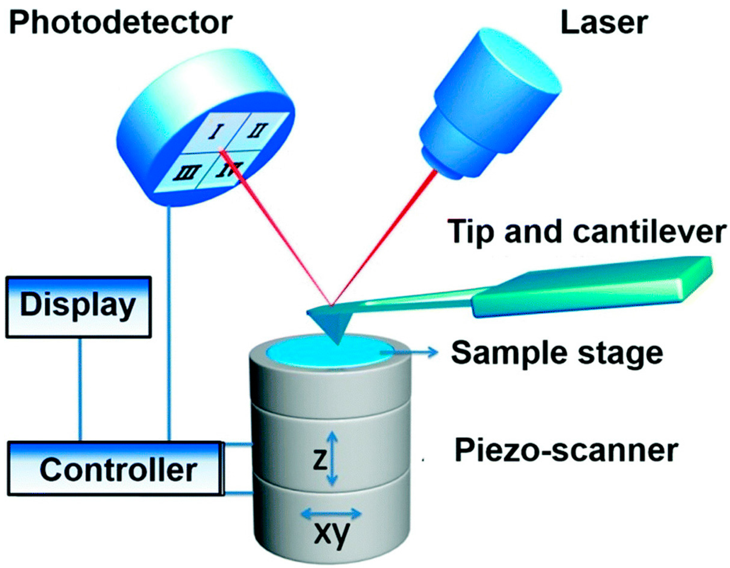
Atomic force microscope diagram. Similarly in atomic force microscopes, depending on the different modes, there is a parameter that serves as the setpoint. For example, in static mode (contact mode) the feedback parameter is cantilever deflection, while in the most common form of tapping mode, the cantilever oscillation amplitude is the feedback parameter. The instrument is ... Nov 16, 2021 · What is a Dissecting microscope (Stereo microscope)? These are also known as stereoscopic microscopes. This is a type of digital optical microscope designed with a low magnification power (5x-250x), by use of light reflected from the surface of the specimen, and not the light reflected the specimen. Atomic force microscope block diagram, laser and photodiodes detection system, second position of the tip, inspired by Atomic force microscope block diagram.png: Date: 21 February 2008: Source: Own work: Author: Twisp: Other versions: Derivative works of this file: Microscopio de fuerza atomica esquema v2.svg diagram shows that solubility of only ~8 wt.% Ag can be achieved at high T in the Cu-rich alloy . Interstitial Solid Solutions: Example • Alloying with smaller atoms (C, N) • Small atoms fill in interstitial spaces • These atoms deform crystal lattice stressand introduce internal Steel is an interstitial iron-carbon alloy Iron: BCC and FCC atomic radii: BCC - 1.24 Å FCC - 1.29 Å ...
Atomic force microscopy (AFM) is a powerful technique that enables the imaging of almost any type of surface, including polymers, ceramics, composites, glass and biological samples. AFM is used to measure and localize many different forces, including adhesion strength, magnetic forces and mechanical properties. File:Atomic force microscope block diagram.svg. Size of this PNG preview of this SVG file: 646 × 599 pixels. Other resolutions: 259 × 240 pixels | 517 × 480 pixels | 647 × 600 pixels | 828 × 768 pixels | 1,104 × 1,024 pixels | 926 × 859 pixels. AFM, which uses a sharp tip to probe the surface features by raster scanning, can image the surface topography with extremely high magnifications, up to 1,000,000X, comparable or even better than electronic microscopes. The measurement of an AFM is made in three dimensions, the horizontal X-Y plane and the vertical Z dimension. §D. Sarid, Scanning Force Microscopy with Applications to Electric, Magnetic and Atomic Forces , Revised Edition, Oxford University Press, 1994. § U. Dürig, "Interaction sensing in dynamic force microscopy", New Journal of
Visualization of Hydration Layers on Muscovite Mica in Aqueous Solution by Frequency-Modulation Atomic Force Microscopy. J. Chem. Phys. 138 (18), 184704. 10.1063/1.4803742 [Google Scholar] Kuchuk K., Sivan U. (2018). Hydration Structure of a Single DNA Molecule Revealed by Frequency-Modulation Atomic Force Microscopy. Noncontact atomic force microscopy (NC-AFM), usually operated in frequency-modulation mode (), has become an important tool in the characterization of nanostructures on the atomic scale.Recently, impressive progress has been made, including atomic resolution with chemical identification and measurement of the magnetic exchange force with atomic resolution (). Atomic force microscopy is arguably the most versatile and powerful microscopy technology for studying samples at nanoscale. It is versatile because an atomic force microscope can not only image in three-dimensional topography, but it also provides various types of surface measurements to the needs of scientists and engineers. 23.05.2019 · (a) Schematic of a graphene membrane before atomic layer deposition (ALD). (b) Optical image of an exfoliated graphene flake with 7 cycles of alumina ALD. (c) Optical image of a pure alumina film after graphene is etched away. (d) Atomic force microscope image of a pressurized 7-cycle pure alumina ALD film with ∆p = 278 kPa. (e) Deflection vs ...
Atomic force microscopy (AFM) is a type of scanning probe microscopy (SPM), with demonstrated resolution on the order of fractions of a nanometer, more than 1000 times better than the optical diffraction limit. The information is gathered by "feeling" or "touching" the surface with a mechanical probe.
schematic diagram of this experiment. Alpha-particles emitted by a 214 83 Bi radioactive source were collimated into a narrow beam by their passage through lead bricks. The beam was allowed to fall on a thin foil of gold of thickness 2.1 × 10 –7 m. The scattered alpha-particles were observed through a rotatable detector consisting of zinc sulphide screen and a microscope. The …
Life Sciences Atomic Force Microscopy (bio - AFM) Atomic force microscopy allows direct visualization of the three-dimensional structure of a sample surface with nanoscale resolution. Unlike many other high-resolution imaging techniques, AFM does not require staining or coating of the sample, leaving native surfaces unaltered.
Atomic Force Microscope (AFM) The atomic force microscope was developed as an alternative to the STM, for use with samples that do not conduct electricity well. The microscope utilizes a cantilever with an extremely sharp probe tip that maintains a constant height above the specimen, typically by direct contact with the sample.
In this article we will discuss about the design of atomic force microscope, explained with the help of a diagram. An atomic force microscope instead of using a lens is provided with a probe to examine the surface of a specimen with a sharp tip which may be several micrometers in and less than 10 nm in diameter at the point near field to be examined. The tip lies at the end of the lever which ...
29.12.2021 · One of the most powerful ways to harness nanoscale flexoelectricity is to press the surface of a material through an atomic force microscope (AFM) tip to generate large strain gradients. This so-called AFM tip pressing allows us to locally break the inversion symmetry in any materials and study all the fascinating physical phenomena associated with inversion …
Basically, there are few of electrons (Olga and Dieter, 2012), which passes through main parts in AFM. Microscope stage is used for moving thin specimen to produce images. Comparison between AFM tip (Maurice et al., 2007), sample holder and force scanning electron microscopy, atomic force microscopy sensor.
Functionalizing the tip of an atomic force microscope with a CO molecule enabled atomic-resolution imaging of single molecules, and measurement of their adsorption geometry and bond-order relations. In addition, by using scanning tunneling microscopy and Kelvin probe force microscopy, the density of the molecular frontier orbitals and the ...
Functionalizing the tip of an atomic force microscope with a CO molecule enabled atomic-resolution imaging of single molecules, and measurement of their adsorption geometry and bond-order relations. In addition, by using scanning tunneling microscopy and Kelvin probe force microscopy, the density of the molecular frontier orbitals and the ...
Keywords: Atomic force microscopy, Protein structure, Amyloid, Disease, Correlative microscopy, NMR. Abstract: Proteins are versatile macromolecules that perform a variety of functions and participate in virtually all cellular processes. The functionality of a protein greatly depends on its structure and alterations may result in the ...
Here, we review the development and recent achievements using high-speed atomic force microscopy (HS-AFM). The HS-AFM is now the only technique to assess structure and dynamics of single molecules, revealing molecular motor action and diffusion dynamics. From this imaging data, watching molecules at work, novel and direct insights could be ...
Abstract. Atomic force microscopy (AFM) with molecule-functionalized tips has emerged as the primary experimental technique for probing the atomic structure of organic molecules on surfaces. Most experiments have been limited to nearly planar aromatic molecules due to difficulties with interpretation of highly distorted AFM images originating ...
24.12.2021 · The atomic force microscope (AFM) is a type of scanning probe microscope whose primary roles include measuring properties such as magnetism, height, friction. The resolution is measured in a nanometer, which is much more accurate and effective than the optical diffraction limit. It uses a probe for measuring and collection of data involves touching …
An atom is the smallest unit of ordinary matter that forms a chemical element.Every solid, liquid, gas, and plasma is composed of neutral or ionized atoms. Atoms are extremely small, typically around 100 picometers across. They are so small that accurately predicting their behavior using classical physics—as if they were tennis balls, for example—is not possible due to quantum …
Atomic absorption spectroscopy (AAS) and atomic emission spectroscopy (AES) is a spectroanalytical procedure for the quantitative determination of chemical elements using the absorption of optical radiation (light) by free atoms in the gaseous state. Atomic absorption spectroscopy is based on absorption of light by free metallic ions.
18.11.2021 · The Atomic Force Microscope (AFM) takes the image of the surface topography of the sample by force by scanning the cantilever over a section of interest. Depending on how raised or how low the surface of the sample is, it determines the deflection of the beam, which is monitored by the Positive-sensitive photo-diode (PSDP). The microscope has a feedback loop …
The transmission electron microscope (TEM) and scanning electron microscope (SEM) are two common forms. Scanning probe microscopy produces images of even greater magnification by measuring feedback from sharp probes that interact with the specimen. Probe microscopes include the scanning tunneling microscope (STM) and the atomic force microscope ...
Nov 20, 2021 · In the past decades, atomic force microscopy (AFM) has significantly deepened our understanding of water-solid interfaces at molecular scale. In this review, we describe the recent progresses on probing water-solid interfaces by noncontact AFM, highlighting the imaging of interfacial water with ultrahigh spatial resolution.
light or electrons, atomic force microscopes use a very fine tip that scan the sample, producing a high resolution image of the surface. Therefore, with light and electron microscopes, internal structure of cells has been possible to analyze. On the opposite, the atomic force microscope is a surface instrument. The approach mentioned in this
Atomic-force microscopy is a reference method for traceable and correlative measurements of nanostructures. The Nanostructure Fabrication and Measurement Group is developing critical-dimension and traceable microscope systems to calibrate probe tips and microscopy standards, and measure diverse devices ranging from waveguides to nanoparticles.
Scanning Probe Microscopy (SPM) ~1600 Light Microscope 1938: Transmission Electron Microscope 1964: Scanning Electron Microscope 1982: Scanning Tunneling Microscope 1984: Scanning Near-field Optical Microscope 1986: Atomic Force Microscope - magnetic force, lateral force, chemical force...
Definition of Atomic Force Microscope (AFM) The AFM, also known as the atomic force microscope (AFM) is a sort scanner probe. Its principal functions include measuring characteristics like height, magnetism and friction. Resolution is determined in millimeter. It is more precise and efficient than the optical Diffraction Limit.
A high-speed atomic force microscope (HS-AFM) requires a specialized set of hardware and software and therefore improving video-rate HS-AFMs for general applications is an ongoing process.
Kelvin probe force microscopy (KPFM), also known as surface potential microscopy, is a noncontact variant of atomic force microscopy (AFM). By raster scanning in the x,y plane the work function of the sample can be locally mapped for correlation with sample features. When there is little or no magnification, this approach can be described as using a scanning Kelvin …
Diagram. Procedure. Adjustment of a travelling microscope. To get a sufficient amount of light, place the travelling microscope (M) near the window. To make the base of the microscope horizontal, adjust the levelling screw. For clear visibility …
To learn more about Atomic Force Microscopy, click through. AFM microscopes are among the best solutions for measuring the nanoscale surface metrology and material properties of samples. A conventional compound light microscope is limited to a maximum sample magnification of approximately 1000x; a quantity that is dictated by the wavelengths of ...
Functionalizing the tip of an atomic force microscope with a CO molecule enabled atomic-resolution imaging of single molecules, and measurement of their adsorption geometry and bond-order relations. In addition, by using scanning tunneling microscopy and Kelvin probe force microscopy, the density of the molecular frontier orbitals and the ...
Visualization of solvation structure on Li 4 Ti 5 O 12 (111)/ ionic liquid-based electrolyte interface by atomic force microscopy Yifan Bao, Mitsunori Kitta, Takashi Ichii et al.-Solvation structure on water-in-salt/mica interfaces and its molality dependence investigated by atomic force microscopy Takashi Ichii, Satoshi Ichikawa, Yuya Yamada ...











0 Response to "38 atomic force microscope diagram"
Post a Comment