36 sheep brain labeled diagram
Game Points. 25. 100% needed. Something different? Weakest Link - Continents of the World 7p Text Game. Can you see the hidden message in these logos? 12p Shape Quiz. The world - Fifteen islands 15p Image Quiz. The Most Misspelled Word In The English Langauge 10p Multiple-Choice. Time Zones of the USA 6p Image Quiz. This map shows the major structures of the sheep brain with an active cursor to help identify the structures. Neuroanatomy Tutorial I: Basic Anatomy of the Brain. Point to any region of this midsaggital sheep brain image (medial view) to highlight that structure. Click the left mouse button to identify the structure you are pointing to.
neuroanatomy by examining the brain of the sheep. We will be looking as several structures within the central nervous system (CNS), which is composed of the brain and spinal cord. You will learn individual structures now; later you will be looking at brain systems to see how different parts of the brain form functional units.
Sheep brain labeled diagram
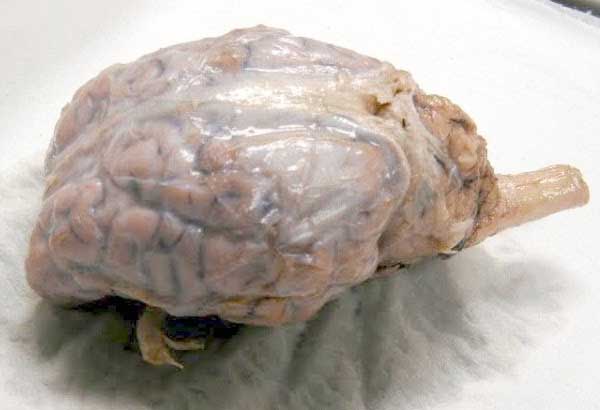
Jun 6, 2018 - A virtual sheep brain dissection guides anatomy studies with photos & blank diagrams. Also shop complete dissection kits: guide, tools & preserved specimen. Sheep Brain Dissection: Internal Anatomy. Place the brain with the curved top side of the cerebrum facing up. Use a scalpel (or sharp, thin knife) to slice through the brain along the center line, starting at the cerebrum and going down through the cerebellum, spinal cord, medulla, and pons. Separate the two halves of the brain and lay them ... The sheep brain is exposed and each of the structures are labeled and described in a sequential manner, in the same way that a real dissection would occur. Sheep Brain Dissection. 1. The sheep brain is enclosed in a tough outer covering called the dura mater. You can still see some structures on the brain before you remove the dura mater.
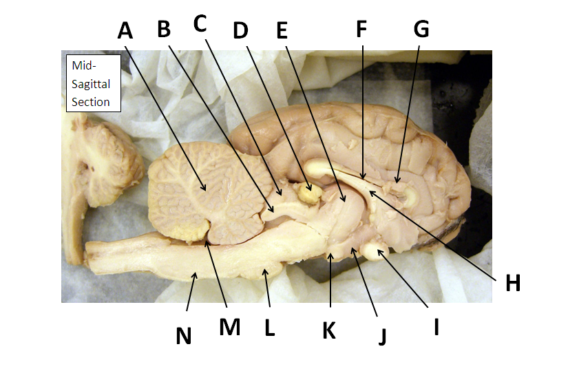
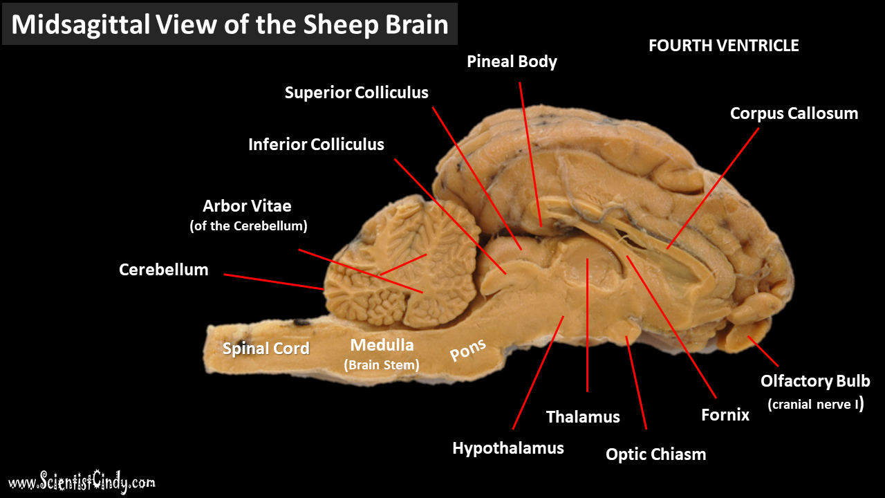
Sheep brain labeled diagram. to anatomy studies. See for yourself what the . cerebrum, cerebellum, spinal cord, gray matter, white matter, and other parts of the brain look like! Observation: External Anatomy . 1. You'll need a . preserved sheep brain. for the dissection. Set the brain down so the flatter side, with the white . spinal cord. at one end, rests on the ... Start studying Sheep Brain Dissection labeled 2. Learn vocabulary, terms, and more with flashcards, games, and other study tools. Instructions - Internal Anatomy: 1. Select a sheep brain that has been cut in half longitudinally (sagitally). Lay the brain flat on the dissecting tray so as to view the internal surface. 2. Identify the internal structures indicated below on the brain. Then label the internal anatomy diagram with all the structures. The rostral colliculus(large arrow label) and the caudal colliculus(small arrow label) together form the tectumof the midbrain. Also labeled are the pineal body(green), the caudate nucleus(1), the floor of the fourth ventricle(white and pink) and cerebellar peduncles(blue = rostral, red = middle, and yellow = caudal). Go Top
To study the structure and function of the mammalian (sheep) brain. References: "Nelson Biology 12" (chapter 9), dissection charts, models, internet, other texts. Instructions - External Anatomy: Obtain a handout with diagrams of the brain from your teacher. Working in groups of two or three, select a sheep brain. Labeled Sheep Brain Diagrams Hol Brain Anatomy Nervous System Anatomy Basic Anatomy And Physiology. Transverse Fissure Cerebral Cortex Human Anatomy And Physiology Brain Anatomy. Sheep Brain External Anatomy Ventral Source By Ntack6076 Hypoglossal Nerve Abducens Nerve Vagus Nerve. This online quiz is called Sheep Brain anatomy, sheep brain Sheep are wonderful and cute. The brain is an interesting organ. It helps with cognition and memory. Almost all the basic task In the body is commanded by the Brain. It is the control center of the body which regulates and control the process crucial for survival Are you interested in learning more about the brain of different animals? Can you answer all the questions of this "Sheep Brain ...
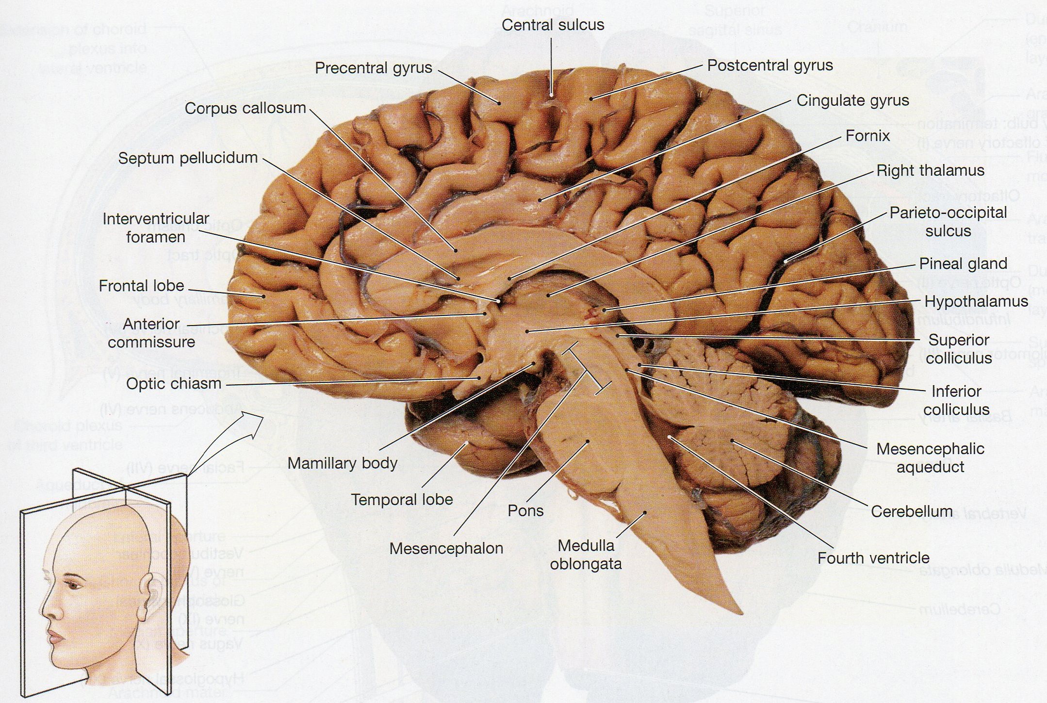
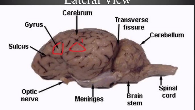
Shows pictures of a sheep and a human brain. Each of the 12 cranial nerves is represented, students color and number each nerve in both brains. Pretty good picture of the sheep brain labeled. Learn the external and internal anatomy of sheep brains with HST's Learning Center science lesson and guide! By the way, concerning Brain Anatomy Worksheet, below we can see particular variation of photos to add more info. brain diagram worksheet, brain diagram and functions worksheet and labeled sheep brain worksheet are three main things we will show you based on the gallery title. Diagram of Sheep Brain - Lateral view Why$dread$a$bump$on$the$head?$ $ October$2012$ Lesson$2:$What$does$thebrain$looklike?$ $ $ $ 2 $ What'supwithallthenewwords?! $ With$all$of$the$new$terminology$in ...
Physiological Psychology
Sheep Brain Neuroanatomy Online Self-Test. Use each diagram as a reference, and selected the correct answer for each lettered structure. You may find it useful to open the diagrams in a separate window to review while answering each question. Dorsal Surface.

sheep brain labeling #2 Diagram | Quizlet
DISSECTION OF THE SHEEP'S BRAIN Introduction The purpose of the sheep brain dissection is to familiarize you with the three-dimensional structure of the brain and teach you one of the great methods of studying the brain: looking at its structure. One of the great truths of studying biology is the saying that "anatomy precedes physiology".

BIO201-Sheep Brain
5 3 11 6 22 16 18 1. Gray Matter 2. White Matter 3. Corpus Callosum 4. Lateral Ventricle 5. Caudate Nucleus 6. Septum Pellucidum 7. Fornix 8.
Sheep Brain Dissection with Labeled Images
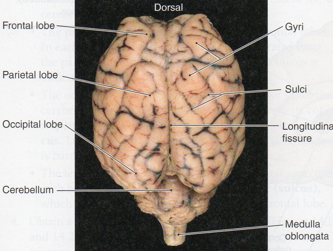
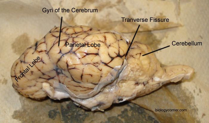
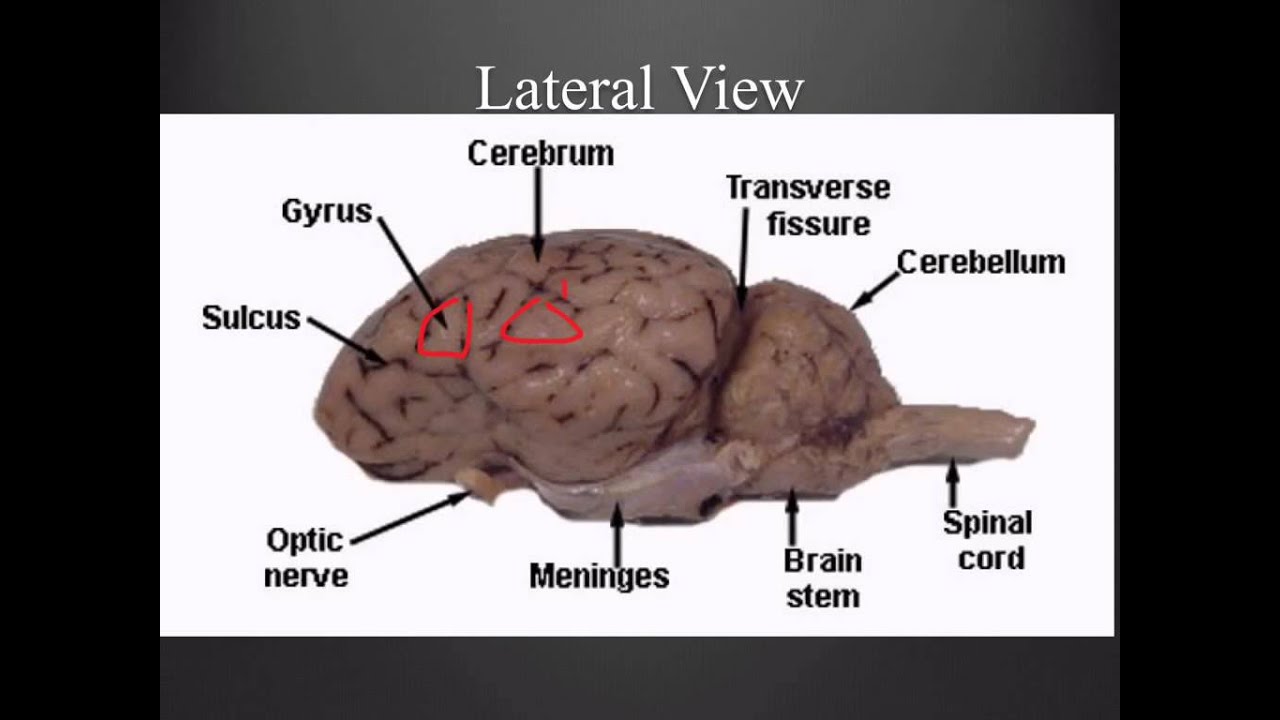
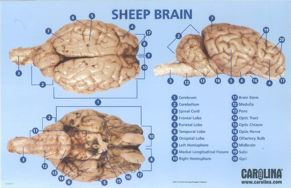
The lobes of the brain are visible, as well as the transverse fissure, which separates the cerebrum from the cerebellum. The convolutions of the brain are also visible as bumps (gyri) and grooves (sulci). Use the diagram below to help you locate these items. Dorsal View of the Sheep Brain . 8.

Sheep Brain Images
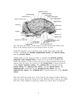
Sheep Neuroanatomy Lab- Labeling Worksheet Psychology 2315- Brain and Behaviour Kwantlen Polytechnic University Figure 1: Dorsal view Cerebellum, Frontal lobe, Occipital lobe, Parietal lobe, and Temporal lobe. Temporal Parietal Lobe Frontal Lobe Cerebellum Occipital Lobe
Sheep Brain Dissection Project Guide | HST Learning Center
AP Biology Sheep Brain Dissection Lab (50 points) Directions: You will be making three (3) detailed drawings of the Sheep Brain in your lab notebook. List the complete scientific classification (domain species) of a sheep. (You will have to look this up on the internet.) DIAGRAM 1: DORSAL VIEW 1.
Index of /files/OCC_VIDEO/upload/Faculty_Resources/acamilo ...
Sheep Brain Anatomy Lab Manual. Based on original material by R. N. Leaton, Dartmouth College. Contributors to this version: Al Sorenson, Lisa Raskin, Sarah Turgeon, Steve George, and JP Baird. I. Introduction. The brain of the sheep is useful for study because its anatomy is similar to human brain anatomy. Although exact proportions (and names ...
Sheep Brain Dissection
2021 2 by Richard Hodgson. Each hemisphere has a frontal temporal parietal and occipital. With more related things such sheep brain diagram labeled brain nervous system worksheet and blank heart diagram. The diagram of the brain is useful for both Class 10 and 12. THE LOBES Occipital lobe Lower back of the brain.
Sheep Brain Neuroanatomy Online Self-Test | KPU.ca - Kwantlen ...
Sheep Brain Dissection Lab Companion In 2021 Brain Anatomy Anatomy And Physiology Brain. Sheep Brain External View Labeled Anatomia Veterinaria Anatomia Veterinaria. Diagram Of The Brain Pictagram Brain Diagram Dissection Brain Anatomy. Biology 02 Steiner Flashcards Sheep Brain Studyblue. Sheep Brain Dissection Project Guide Hst Learning Center ...

Sheep brain | Atlas of Comparative Vertebrate Anatomy
Start studying Sheep Brain Dissection labeled. Learn vocabulary, terms, and more with flashcards, games, and other study tools.

Resources for Teaching Mammalian Neuroanatomy Using Sheep ...
function, and pathology. Those students participating in Sheep Brain Dissections will have the opportunity to dissect and compare anatomical structures. At the end of this document, you will find anatomical diagrams, vocabulary review, and pre/post tests for your students. The following topics will be covered: 1.
Sheep Brain Explora on Guide
The sheep brain is exposed and each of the structures are labeled and described in a sequential manner, in the same way that a real dissection would occur. Sheep Brain Dissection. 1. The sheep brain is enclosed in a tough outer covering called the dura mater. You can still see some structures on the brain before you remove the dura mater.
Sheep Brain Dissection Guide
Sheep Brain Dissection: Internal Anatomy. Place the brain with the curved top side of the cerebrum facing up. Use a scalpel (or sharp, thin knife) to slice through the brain along the center line, starting at the cerebrum and going down through the cerebellum, spinal cord, medulla, and pons. Separate the two halves of the brain and lay them ...
External sheep brain dissection guide
Jun 6, 2018 - A virtual sheep brain dissection guides anatomy studies with photos & blank diagrams. Also shop complete dissection kits: guide, tools & preserved specimen.
Sheep Brain Dissection
sheep brain anatomy
11c Brain Anatomy
Sheep Brain Dissection with Labeled Images
The Brain - SCIENTIST CINDY
Sheep brain dissection - Bisc 163 - StuDocu

inferior view of sheep brain

Sheep Brain Dissection | Carolina.com
Sheep Brain Dissection

Sheep brain | Atlas of Comparative Vertebrate Anatomy
Physiological Psychology

Dissecting Sheep Brains With Sixth Graders | Brains Explained

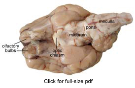
Diagram of Sheep Brain - Lateral view

Sheep Brain Images

Sheep Brain - Animal Physiology
Sheep Brain Dissection
Medical Detectives Lesson 27

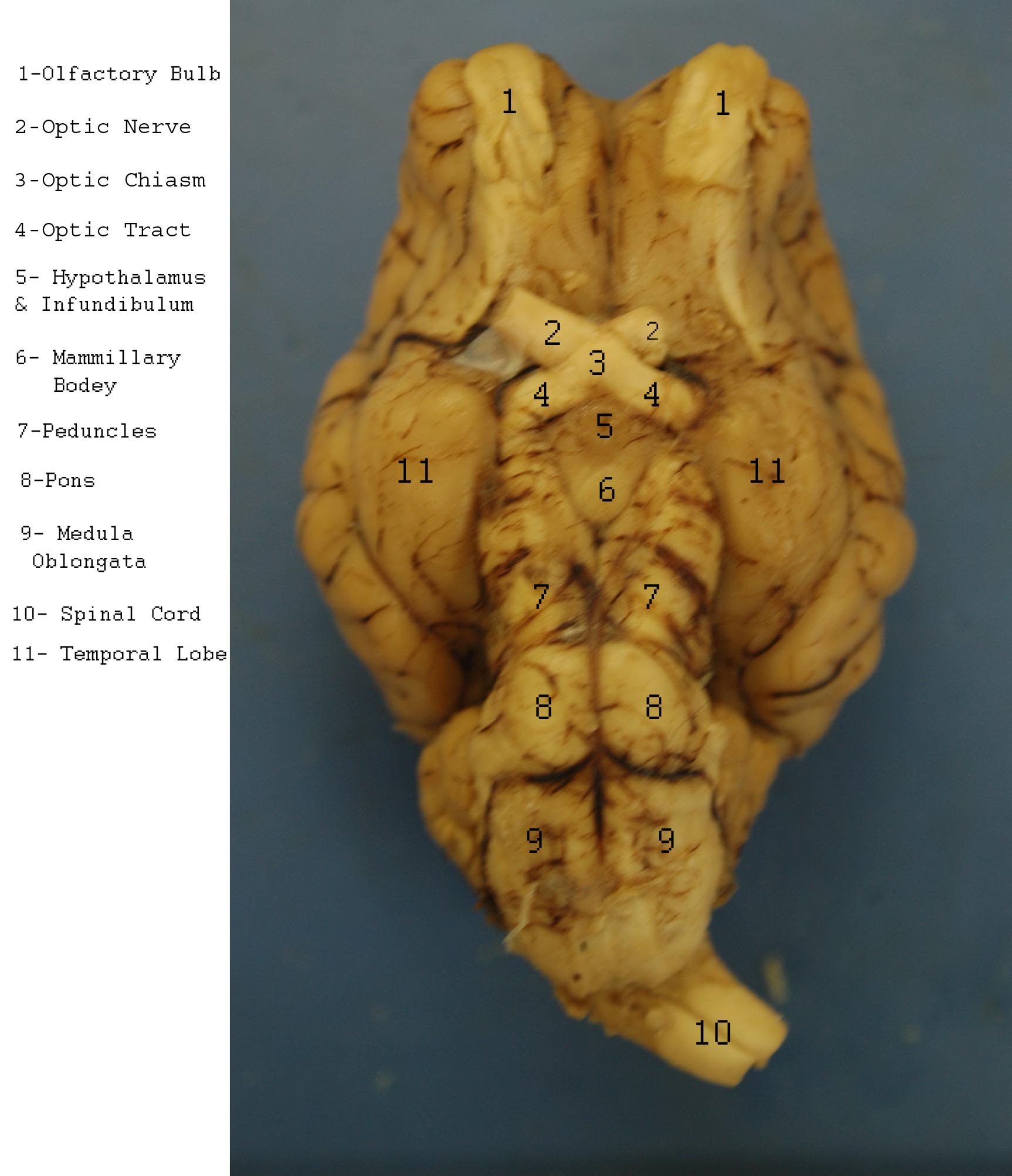
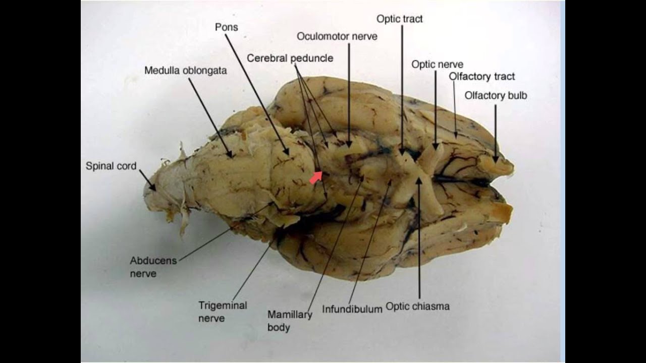
Sheep brain - external anatomy (ventral) | Hypoglossal nerve ...

Diagram of Sheep Brain - Inferior view

Sagittal Section of Sheep Brain Quiz
Sheep Brain Dissection Guide - YouTube
Untitled
0 Response to "36 sheep brain labeled diagram"
Post a Comment