36 identify the structures labeled a, b, and c in the diagram of a sarcomere above.
11. Label the thick and thin filaments in Figs. A, B, and C above. 12. There are three sarcomeres shown in the diagram below. a) In Sarcomere 1, identify the location within the sarcomere of the cross section indicated by Figure A in Model 3. Draw a vertical line and label it A. b) In Sarcomere 2, identify the location within the sarcomere of the cross 23) Identify the specific structure indicated by Label A. A) Myosatellite cell. B) Myoblast. C) Schwann cell. D) Motor end plate. E) Motor neuron. D. 24) Identify the specific structure indicated by Label C. A) Dendrite.
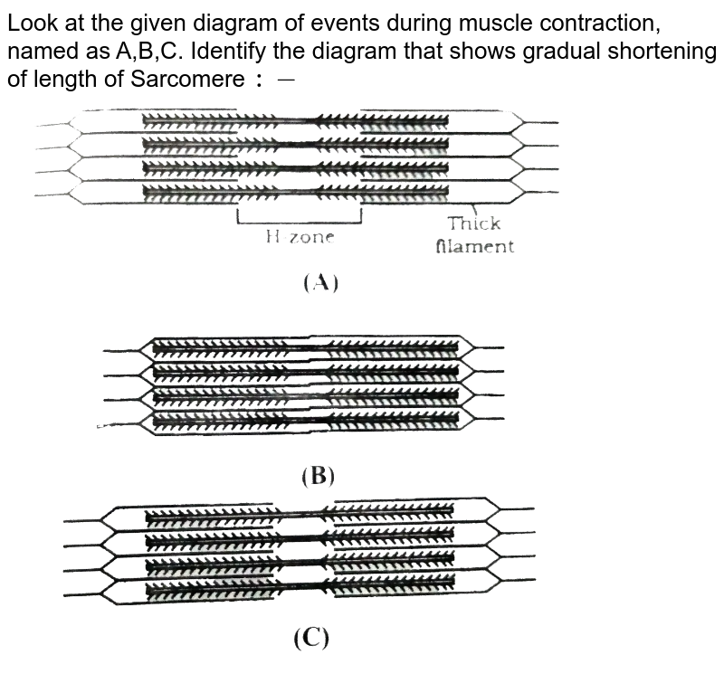
The diagrams in Model 3 are cross sections of a sarcomere that show the filaments at various locations within a sarcomere. thick thin Fig. A 10. Label the thick and thin filaments in Figs. A, B, and C above. 11. In the diagram below, draw three vertical lines showing the locations within a
Identify the structures labeled a, b, and c in the diagram of a sarcomere above.
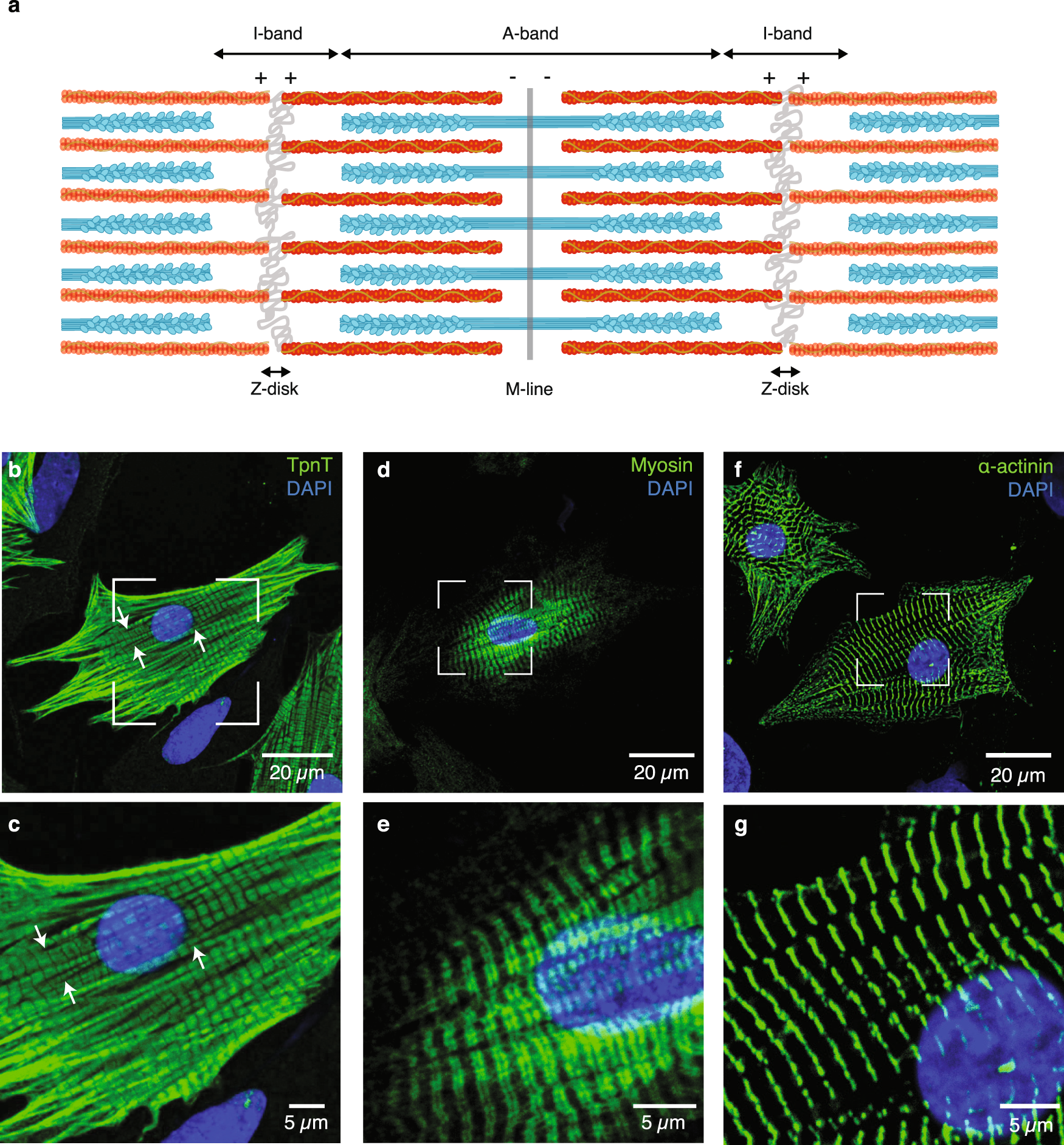
A sarcomere is defined as the region of a myofibril contained between two cytoskeletal structures called Z-discs (also called Z-lines), and the striated appearance of skeletal muscle fibers is due to the arrangement of the thick and thin myofilaments within each sarcomere (Figure 10.2.2). A sarcomere is the functional unit of striated muscle. This means it is the most basic unit that makes up our skeletal muscle. Skeletal muscle is the muscle type that initiates all of our voluntary movement. Herein lies the sarcomere's main purpose. Sarcomeres are able to initiate large, sweeping movement by contracting in unison. A Fig. B Fig. QUESTIONS: 10. Label the thick and thin filaments in Figs. A, B, and C above 11. There are three sarcomeres shown in the diagram below. Sarcomere 1 Sarcomere 2 Sarcomere 3 a) In Sarcomere 1, identify the location within the sarcomere of the cross section indicated by Figure A in Model 3.
Identify the structures labeled a, b, and c in the diagram of a sarcomere above.. The diagrams in Model 3 are cross sections of a sarcomere that show the filaments at various locations within a sarcomere. 10. Label the thick and thin filaments in Figs. A, B, and C above. 11. In the diagram below, draw three vertical lines showing the locations within a sarcomere of the cross sections indicated by Figures A, B, and C. Label each C) Tendons function to move various parts of the skeleton in response to skeletal muscle contraction. D) One end of the tendon is usually linked to bone and the other to a skeletal muscle. E) None of the above. Answer: B 2. The contractile unit of a muscle is: A) muscle fibre B) myofilament (myofibril) C) sarcomere D) myosin E) actin Answer: C 3. Recognize a sarcomere as a complete contractile unit. Relate the structure of the sarcomere to the distribution of actin and myosin within the myofibril. Describe the sliding-filament hypothesis of muscle contraction. Identify the role of calcium ions (Ca2+), the troponin-tropomyosin complex, and ATP. Recognize that Draw a vertical line and label it A. b) In Sarcomere 2, identify the location within the sarcomere of the cross section indicated by Figure B in Model 3. Draw a vertical line and label it B. c) In Sarcomere 3, identify the location within the sarcomere of the cross section indicated by Figure C in Model 3. Draw a vertical line and label it C. Model 2 Muscle Contraction 13. Which of the figures (A, B, or C) represents a cross section in the H zone? B 14.
(ii) Explain how these structures help in the absorption of substances from the small intestine. (1) (b) (i) The scale bar on this drawing represents a length of 0.1μm. Which of the labeled structures on the diagram holds muscles with similar functions together, allows free movement of muscles, and fills spaces between muscles? ... In the figure above, what letter correctly identifies tropomyosin? B. ... which structure helps return a stretched sarcomere to its resting length? C. A, B, and C above. 2. In the diagram below, draw three vertical lines showing the locations within a sarcomere of the cross sections indicated by Figures A, B, and C. Label each of the lines. A B thin thin thick thick C A B C There are three sarcomeres shown in the diagram ft Sarcomere 1 Sarcomere 2 Sarcomere a) In Sarcomere 1, identify the location within the sarcomere of the cross indicated by Figure A in Model 3. Drawa vertical line and at various oo A B o C C above. below. 3 section label it A. 6)
Label the thick and thin filaments in Figs. A, B, and C above. 12. There are three sarcomeres shown in the diagram below. Sarcomere 1 Sarcomere 2 Sarcomere 3 a) In Sarcomere 1, identify the location within the sarcomere of the cross section indicated by Figure A in Model 3. various locations within a sarcomere. 1. Label the thick and thin filaments in Figs. A, B, and C above. 2. In the diagram below, draw three vertical lines showing the locations within a sarcomere of the cross sections indicated by Figures A, B, and C. Label each of the lines. 3. Which of the figures (A, B, or C) represents a cross section in the H zone? 4. Identify the structures labeled A, B, and C in the diagram of a sarcomere above. A = Actin filament; B = Myosin filament; C = Titin The sliding filament theory states that during muscle contraction Sarcomere 1 Sarcomere 2 Sarcomere 3 a) In Sarcomere 1, identify the location within the sarcomere of the cross section indicated by Figure A in Model 3. Draw a vertical line and label it A. b) In Sarcomere 2, identify the location within the sarcomere of the cross section indicated by Figure B in Model 3. Draw a vertical line and label it B.
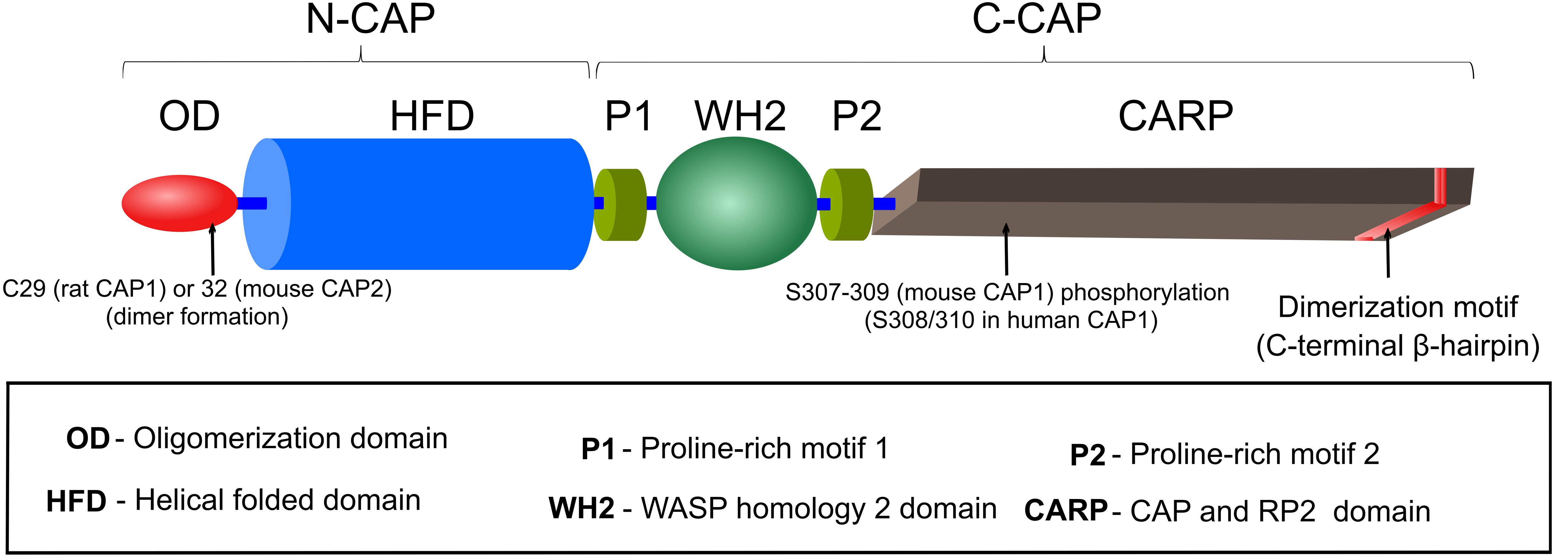
Frontiers | CAPt'n of Actin Dynamics: Recent Advances in the ...
Sarcomere 1 Sarcomere 2 Sarcomere 3 a) In Sarcomere 1, identify the location within the sarcomere of the cross section indicated by Figure A in Model 3. Draw a vertical line and label it A. b) In Sarcomere 2, identify the location within the sarcomere of the cross section indicated by Figure B in Model 3. Draw a vertical line and label it B. A B C Fig. A Fig. B Fig. C Thick
The giant protein titin regulates the length of the striated ...
Start studying UNIT 5: Label the parts of the Sarcomere. Learn vocabulary, terms, and more with flashcards, games, and other study tools.
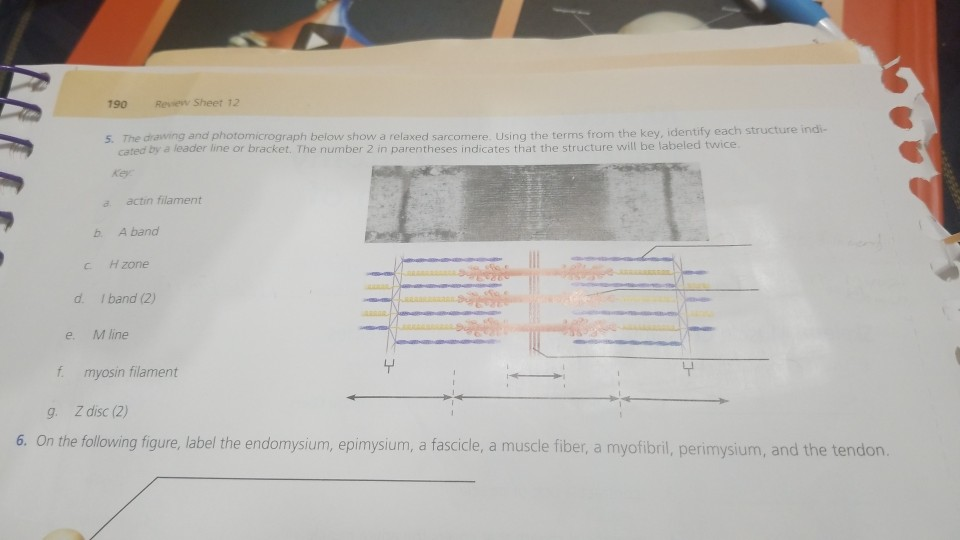
Solved 190 Review Sheet 12 identify each structure indi- 5 ...
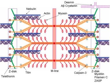
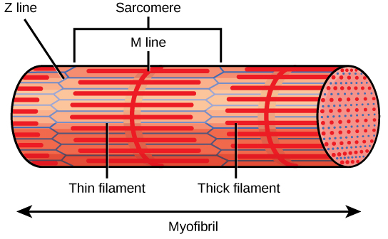
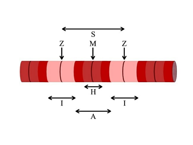
Sarcomere H zone Thin (actin) filament Thick (myosin) filament Z disc Z disc M line (c) Small part of one myofibril enlarged to show the myofilaments responsible for the banding pattern. Each sarcomere extends from one Z disc to the next.
Muscles COntinued
Label the thick horizontal filament THICK filament. 2. Label the thin horizontal filament THIN filament. 3. How many sarcomeres are shown in the above model? 4. Based on your observations of the location of thick and thin filaments, describe each of the following: a) A band b) I band c) H zone d) Z disc e) M line 5.

A) Illustration of skeletal muscle structure copied with ...
Mar 23, 2018 · Identify the structures labeled a b and c in the diagram of a sarcomere above. Indicated by figure a in model 3. Skeletal Muscle Anatomy And Physiology I B myosin filament. Identify the structures labeled a b and c in the diagram of a sarcomere above. Draw a vertical line and label it a. A actin filament. A at the beginning of a contraction.

Study Guide Flashcards | Quizlet
Identify the structures labeled A, B, and C in the diagram of a sarcomere above. A is a "blue" filament, attached to the Z line at the ends of a sarcomere. B is a "red" filament, crossing the M band. C is a "grey" filament attached to both B and the Z line.

Skeletal MyBP-C isoforms tune the molecular contractility of ...
The given below diagram represents the gastric glands. Label it from A to D and choose the correct option accordingly. (a) ... Identify A, B, C and D in the given diagram and choose the correct combination. (a) A-Thyroid, B-Trachea, C-Vocal cord, D-Parathyroid glands ... (b) sarcomere (c) myofibrils similar in length and diameter (d) All of the ...

A&P Module 6 Flashcards | Quizlet
Jan 23, 2019 · A sarcomere is the basic unit of striated muscle tissue. It is the repeating unit between two Z lines. Skeletal muscles are composed of tubular muscle cells which. Sarcomeres are composed of thick filaments and thin filaments. The thin filaments Look at the diagram above and realize what happens as a muscle contracts.

Myogenic Stage, Sarcomere Length, and Protease Activity ...
(b) Simplified diagram of a myosin II molecule; a long rod on the end of which are two myosin heads which bind and hydrolyse ATP and bind actin. Myosin molecules (b) aggregate to form myosin filaments (c); the rods are in the backbone and the heads are almost helically arranged on the filament surface. The head ends of the rods, myosin S2 ...

Cardiac Muscle Tissue | Boundless Anatomy and Physiology
A Fig. B Fig. QUESTIONS: 10. Label the thick and thin filaments in Figs. A, B, and C above 11. There are three sarcomeres shown in the diagram below. Sarcomere 1 Sarcomere 2 Sarcomere 3 a) In Sarcomere 1, identify the location within the sarcomere of the cross section indicated by Figure A in Model 3.

Sarcomeres | BioNinja
A sarcomere is the functional unit of striated muscle. This means it is the most basic unit that makes up our skeletal muscle. Skeletal muscle is the muscle type that initiates all of our voluntary movement. Herein lies the sarcomere's main purpose. Sarcomeres are able to initiate large, sweeping movement by contracting in unison.
Muscular Dystrophy: Practice Essentials, Pathophysiology ...
A sarcomere is defined as the region of a myofibril contained between two cytoskeletal structures called Z-discs (also called Z-lines), and the striated appearance of skeletal muscle fibers is due to the arrangement of the thick and thin myofilaments within each sarcomere (Figure 10.2.2).

Muscle tissue - Knowledge @ AMBOSS

A&P Lab Review Flashcards | Quizlet
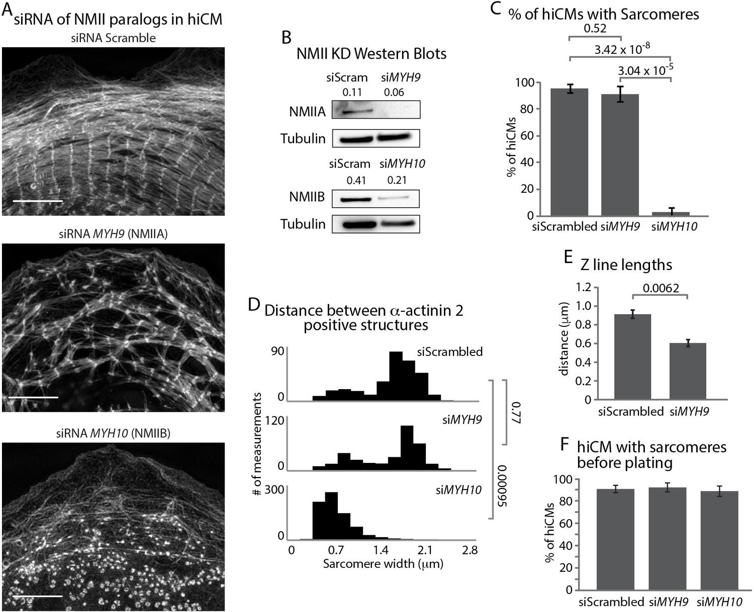
Muscle-specific stress fibers give rise to sarcomeres in ...

How Muscle Structure and Composition Influence Meat and Flesh ...

Site-specific phosphorylation of myosin binding protein-C ...
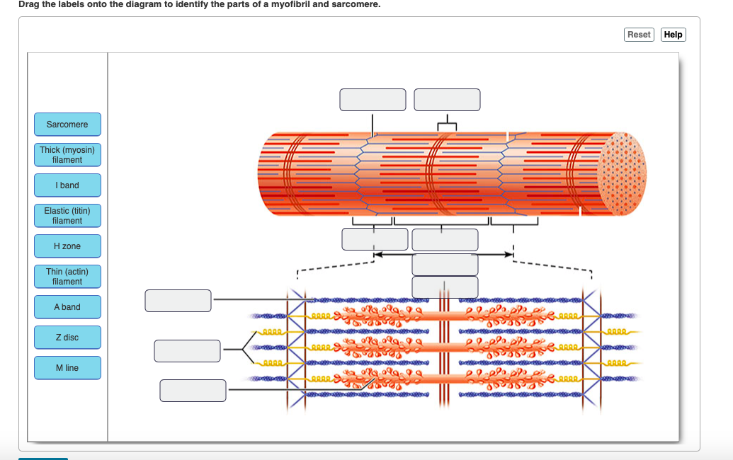
Solved Drag the labels onto the diagram to identify the ...

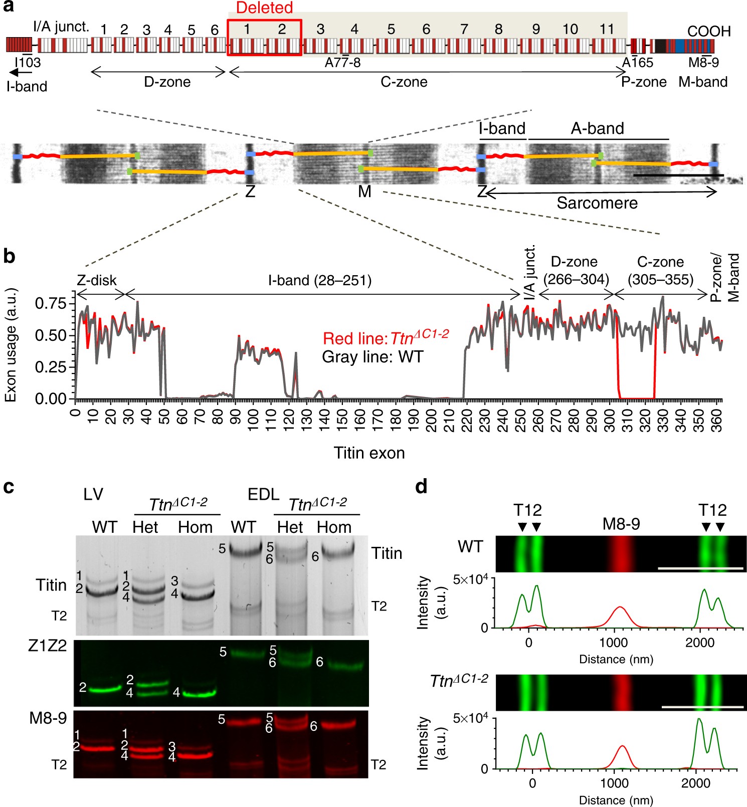
Effects of deleting C-zone repeats on A-band width. a Left ...
Sarcomere - Wikipedia
19.4 Muscle Contraction and Locomotion – Concepts of Biology ...

Lab Muscle Quiz Flashcards | Quizlet
Molecular-scale visualization of sarcomere contraction within ...
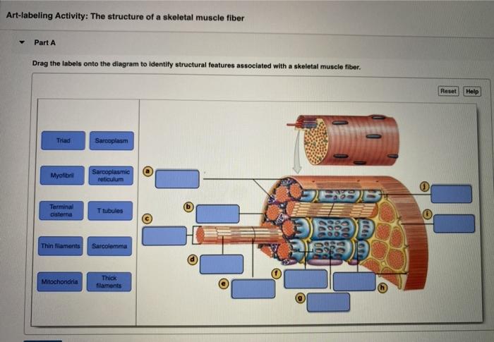
Solved Art-labeling Activity: The Structure of a Sarcomere ...

Chapter 10 Images Flashcards | Quizlet

Ch 13 lab Map, Ch 12 lab map, CH 11 Lab MAP, Ch 10 lab map ...

multi choice chapter 10. Muscle Tissue Flashcards - Easy ...
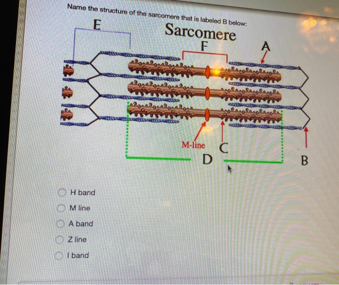
Solved Name the structure of the sarcomere that is labeled B ...
Sarcomere - Definition, Structure, Function and Quiz ...

Cardiac Disorders and Pathophysiology of Sarcomeric Proteins ...

Ch 13 lab Map, Ch 12 lab map, CH 11 Lab MAP, Ch 10 lab map ...

26 Identify The Structures Labeled A, B, And C In The Diagram ...

Solved Art-labeling Activity: Sarcomere Structure Drag the ...

Given below is the figure of a sarcomere. Identify the parts ...
multi choice chapter 10. Muscle Tissue Flashcards - Easy ...
Consder the diagram showing muscle structure below.
0 Response to "36 identify the structures labeled a, b, and c in the diagram of a sarcomere above."
Post a Comment