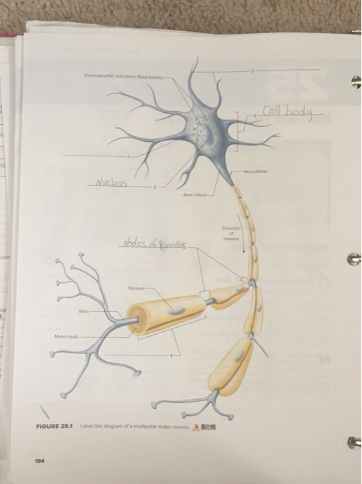
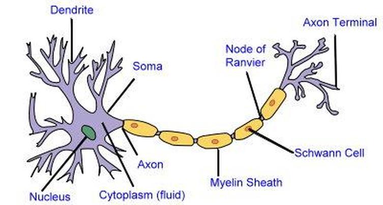
40 labeled diagram of neuron
15/12/2021 · In addition, calcium imaging of MAP2–GcaMP5-labeled neurons indicated active neuronal signaling, confirming that mature and functional DA networks were present in silk-VM organoids (Fig. 8m, n). Diagram Of Neuron with Labels · Dendrites–A branch-like structure that functions by receiving messages from other neurons and allow the transmission of messages ...Cell Membrane Definition: Opening and Closing ...Placenta Definition: Introns and ExonsFermentation Definition: Meiosis DefinitionMitochondria Meaning: Rain Water Harvesting ...
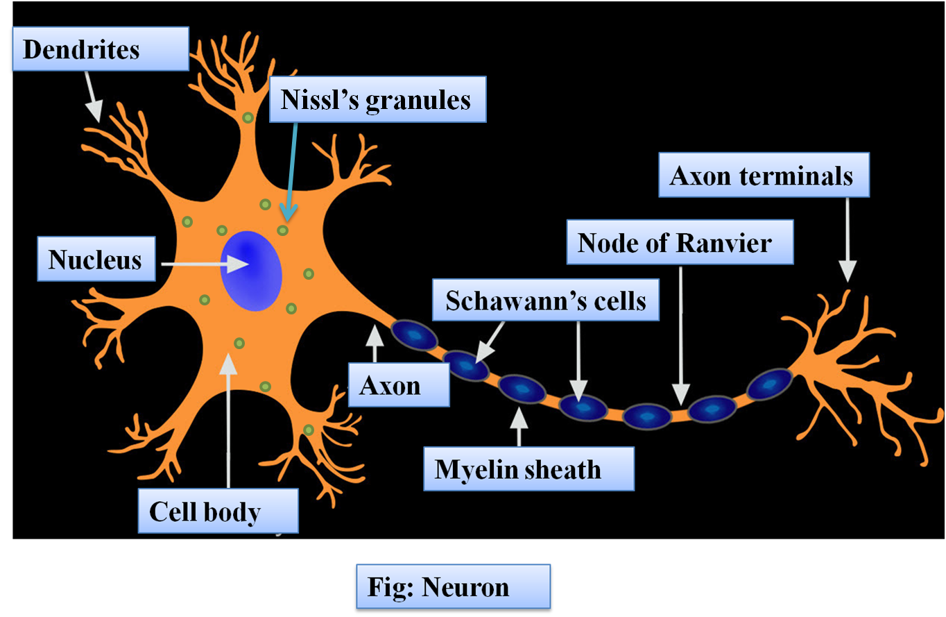
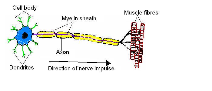
1 answerThe structure of neuron: Nerve cells or neurons are the structural and functional units of the nervous system. It consists of three major parts namely, ...

Labeled diagram of neuron
how to draw structure of neuron/neuron diagram labelled/diagram of neuron/neuron cell. Please watch: "cell structure and functions / animal cell vs plant cell / ... A picture or diagram of a neuron (see the picture below or go to: more about neurons.) If you would like to use this "build a neuron" as a classroom activity, here is a lesson ready to go. Need some play dough but don't have any? Here is a recipe that you can use to make your own: Mix: 1 cup flour, 1/2 cup salt & 2 teaspoon cream of tartar The resting membrane potential of a neuron averages -70mV (millivolts). All neural activities begin with a change in the resting membrane potential of a neuron. The resting membrane potential is maintained by Na+-K+ pumps that actively transport K+ into and Na+ out of the cell. The concentration of Na+ is higher outside than inside the cell. The membrane is more …
Labeled diagram of neuron. Illustration about Labeled diagram of the neuron, nerve cell that is the main part of the nervous system. Illustration of anatomical, peripheral, ... Illustration of Labeled diagram of the neuron, nerve cell that is the main part of the nervous system. vector art, clipart and stock vectors. Find neuron labeled stock images in HD and millions of other royalty-free ... See neuron labeled stock video clips ... Nerve Cell Neuron Labeled Diagram. 20/12/2021 · (N) The fraction of EGFP-labeled (green), mCherry-labeled (red), and dually labeled (yellow) neurons in the Sp5C as a function of bregma, showing that spinal-projecting and LPB-projecting Cbln2 + neurons are largely segregated. Scale bars are labeled in the graphs.
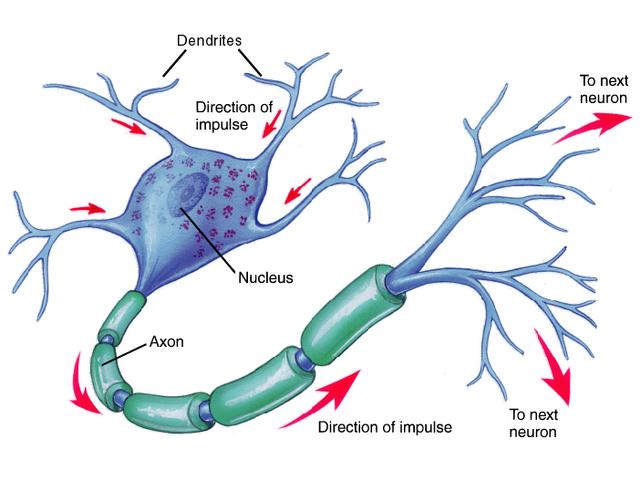
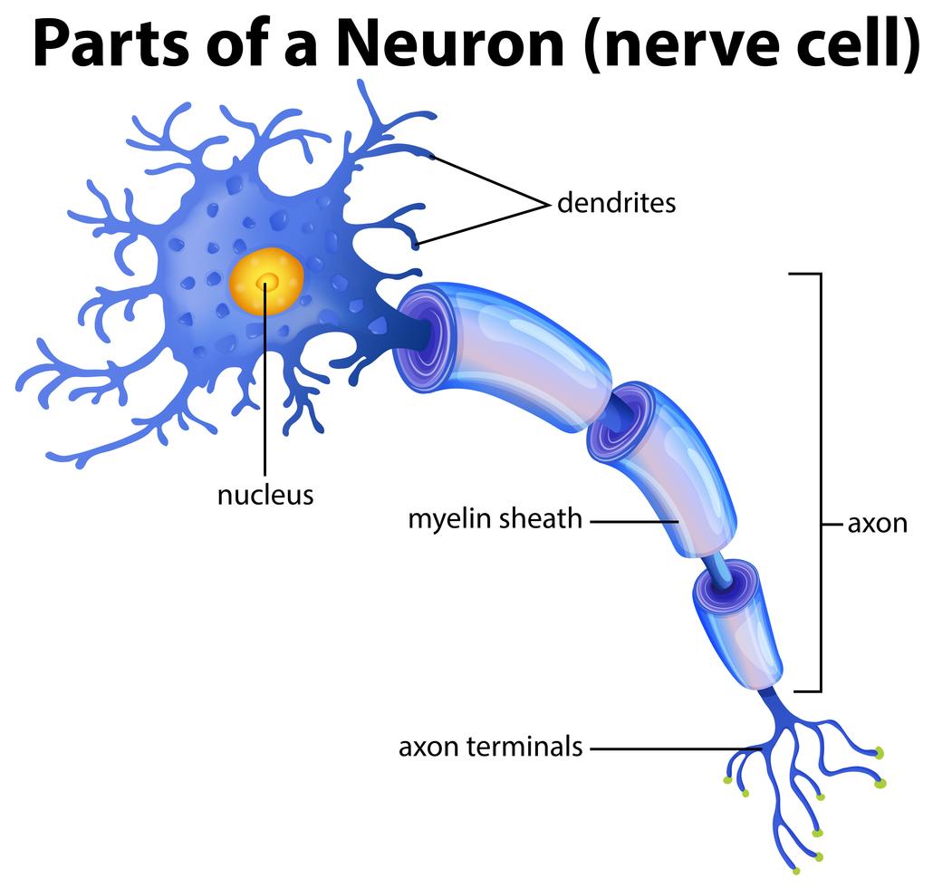
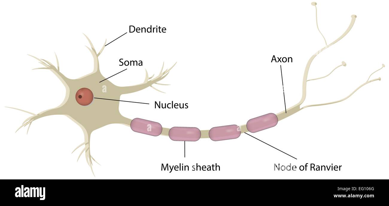
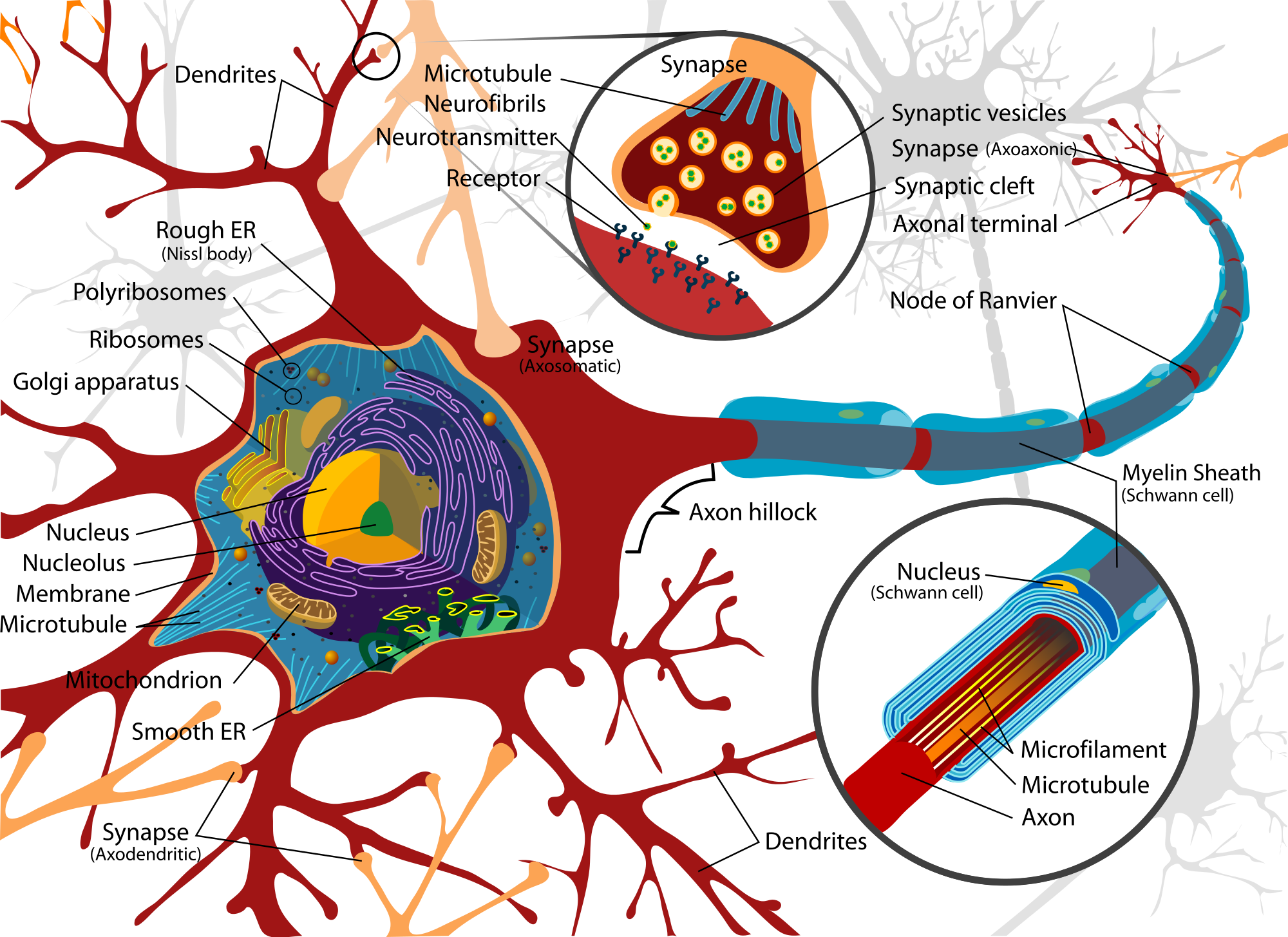
28/06/2020 · A bipolar neuron has the same structures as all neurons but is arranged differently. The central cell body makes a border between the dendrite end and the axon and terminal end. Dendrites are multiple and sometimes it is difficult to know which end is which in a simplified bipolar neuron diagram. Usually, the rounded shape of the terminal ... Draw a labelled diagram of the neuron and describe the structure of the neuron in detail. · 1. Cell body: It forms the cytoplasm of the nerve cell. It is ...1 answer · Top answer: Hint: The cell forms the basic unit of life. Every part of the body has a specialized cell which will perform only a particular activity. They work ... English: Complete neuron cell diagram. Neurons (also known as neurones and nerve cells) are electrically excitable cells in the nervous system that process and transmit information. In vertebrate animals, neurons are the core components of the brain, spinal cord and peripheral nerves. Date: 12 July 2007: Source: Own work. Image renamed from Image:Complete neuron … 06/03/2019 · Grid cells are labeled with the classification they most support. ... et al use t-SNE to make more diverse neuron visualizations, generating diverse starting points for the optimization process by clustering images in the t-SNE map. This reveals a broader picture of what the neuron detects but is still focused on individual neurons. In this article we introduce activation atlases …
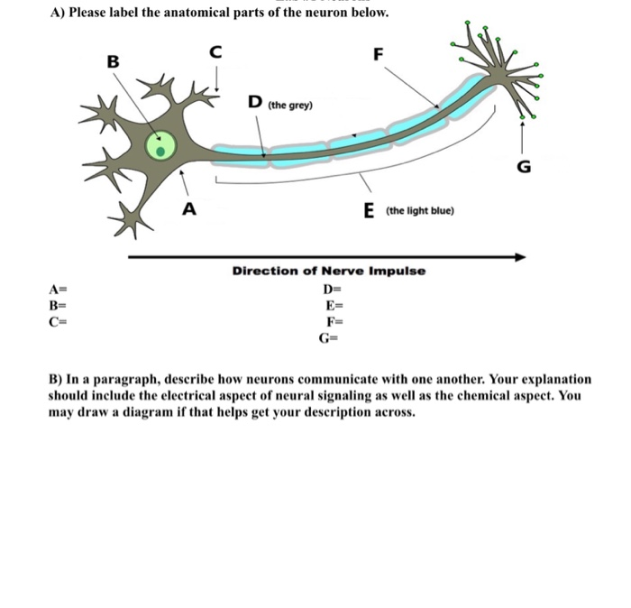
In part 1, students label a diagram of a neuron with structure and function (use Make a Mad, Mad, Mad Neuron as a source). In part 2, they describe what is happening in a labeled diagram of a synapse (use Crossing the Divide as a source). Use as worksheets for students to fill in as they explore the multimedia sources linked below. They may be used as a formative or … Introduction to neurons and glia. How the structure of a neuron allows it to receive and transmit information. 01/12/2021 · Animal cell size and shape. Animal cells come in all kinds of shapes and sizes, with their size ranging from a few millimeters to micrometers. The largest animal cell is the ostrich egg which has a 5-inch diameter, weighing about 1.2-1.4 kg and the smallest animal cells are neurons of about 100 microns in diameter. The resting membrane potential of a neuron averages -70mV (millivolts). All neural activities begin with a change in the resting membrane potential of a neuron. The resting membrane potential is maintained by Na+-K+ pumps that actively transport K+ into and Na+ out of the cell. The concentration of Na+ is higher outside than inside the cell. The membrane is more …
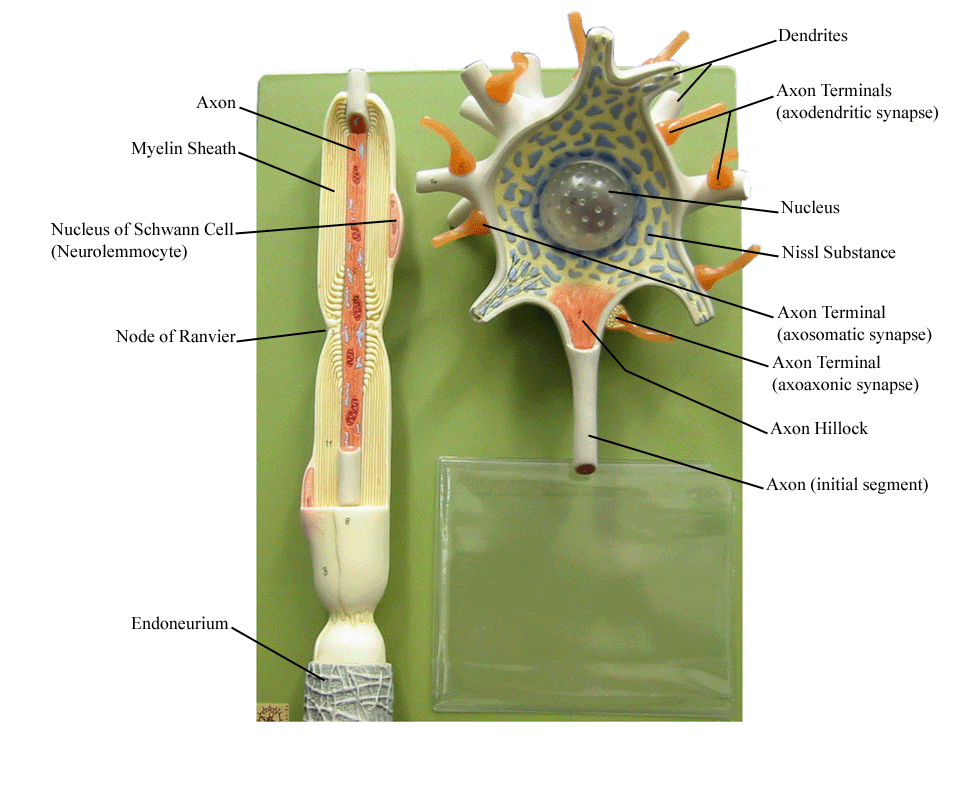
Ultrastructure of Nerves - Classification - Neurones ...
A picture or diagram of a neuron (see the picture below or go to: more about neurons.) If you would like to use this "build a neuron" as a classroom activity, here is a lesson ready to go. Need some play dough but don't have any? Here is a recipe that you can use to make your own: Mix: 1 cup flour, 1/2 cup salt & 2 teaspoon cream of tartar
Neuron Models
how to draw structure of neuron/neuron diagram labelled/diagram of neuron/neuron cell. Please watch: "cell structure and functions / animal cell vs plant cell / ...
Neuron Labeled Diagram - ClipArt Best
35 Neuron Labeled Diagram - Wiring Diagram Database

Image from page 380 of "The Biological bulletin"

Nervous Tissue Mediates Perception and Response | Anatomy ...

Neuron Diagram Labeled - Human Anatomy
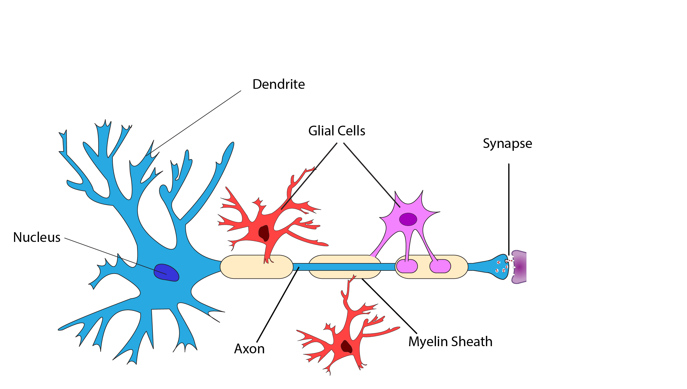
Diagram Of A Neuron With Labels - General Wiring Diagram

Biology: Neurons Structure and Info + Worksheet

Auditory on a diagram of a Neuron - YouTube

12.2 Nervous Tissue - Anatomy & Physiology
Describe the structure of a neuron with the help of class ...

Image from page 374 of "The Biological bulletin"
Neuron Diagram Labeled Axon Hillock - Aflam-Neeeak

Picture Of A Neuron Labeled New Chapter 7 | Cell diagram ...
how to draw structure of neuron/neuron diagram labelled ...
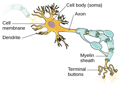
Cells of the Nervous System | Introduction to Psychology
Part of a Neuron Diagram 299698 Vector Art at Vecteezy
place to be

Motor Neuron Detailed And Accurate Labeled Stock ...

Types of Neuron, labeled diagram. Poster | Zazzle

Neuron Cell Diagram Posters by bestsellingts
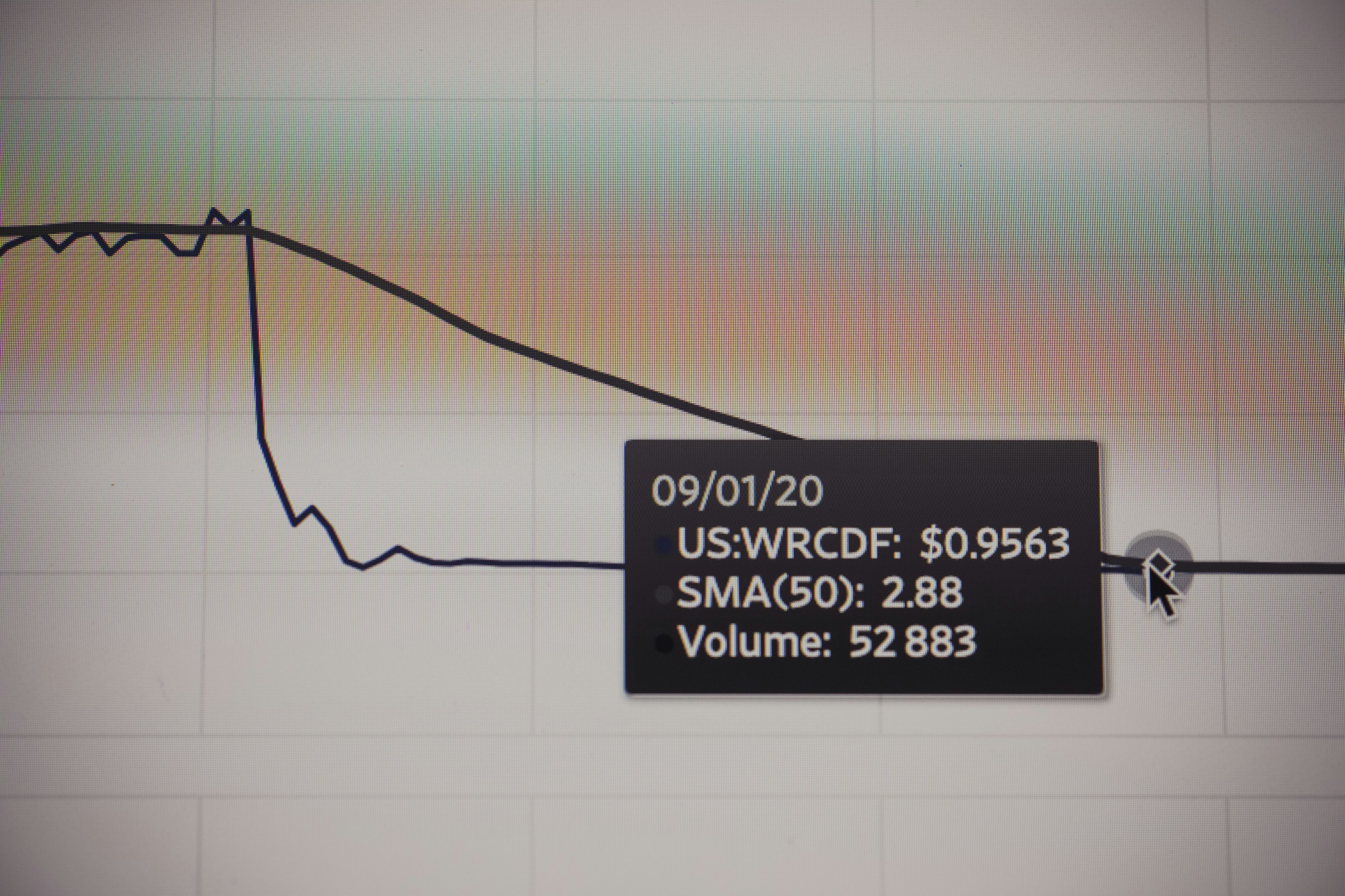
Stock market chart value. Made with analog vintage lens, Leica APO Macro Elmarit-R 2.8 100mm (Year: 1993)
Draw a well labelled diagram of neuron (nerve cells ...

Labeled Neuron Firing Diagram - Aflam-Neeeak
Neuron Labeled Diagram Stock Vector Image & Art - Alamy

Pinned map of the United States of America
The Anatomy and Physiology of Animals/Nervous System ...
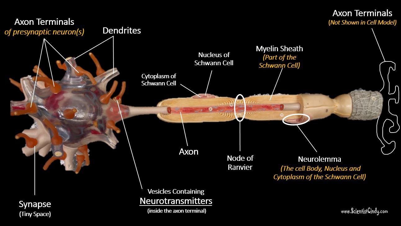
Nervous Tissue - SCIENTIST CINDY
30 Neuron Label Worksheet - Labels For Your Ideas
Nerve Cell Diagram Labeled - ClipArt Best
2D labelled diagram - Nerve cell
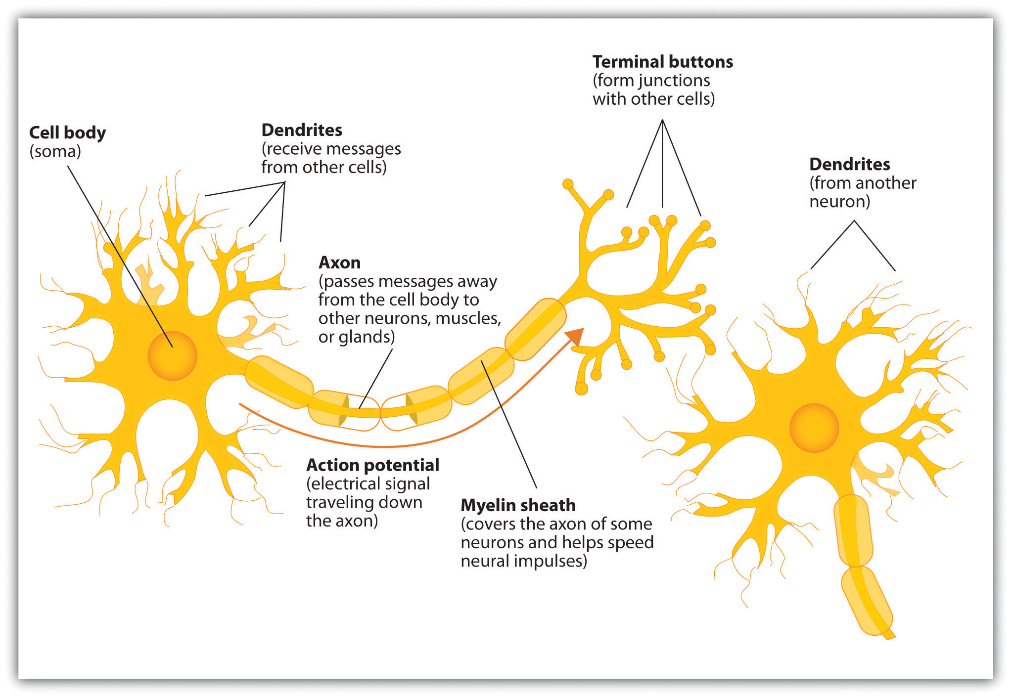
The Neuron Is the Building Block of the Nervous System

draw a labelled diagram of a neuron - Brainly.in

Neuron PowerPoint Diagram

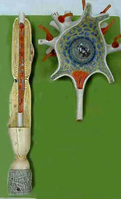
Closeup of skeleton pelvic model
Neuron Diagram Labeled - ClipArt Best

How Do Memories Form? Our Brains May Be More Powerful Than ...
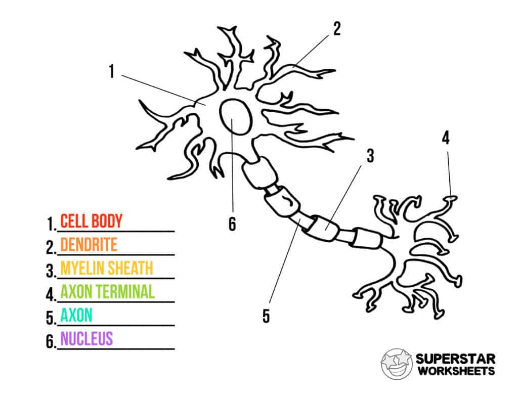
Neuron Cell Worksheets - Superstar Worksheets

draw a labbeled diagram of neuron - Brainly.in
0 Response to "40 labeled diagram of neuron"
Post a Comment