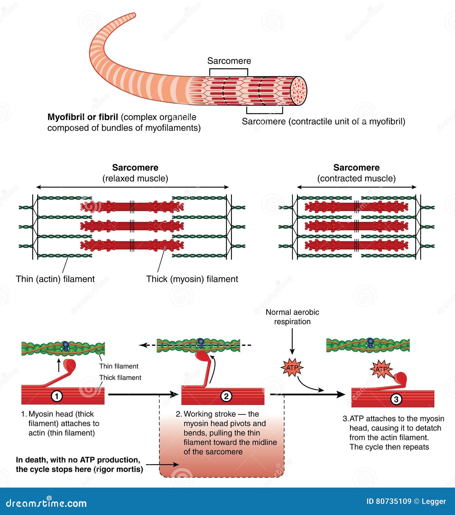
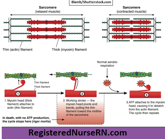
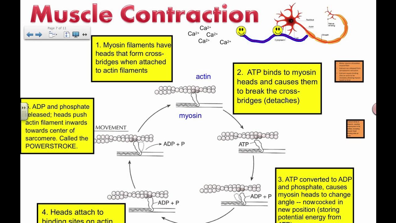
39 diagram of muscle contraction
Muscles of the larynx. There are many muscles that either make up a certain part of the laryngeal structure inside the neck, or that sit adjacent to it and aid in its function.These muscles produce the movements of the larynx and its cartilages, thus enabling the proper air conduction, speech, movements of the epiglottis and airways protection. The muscles of the larynx are divided into two ... Anatomy Structure . Within the gluteus maximus, fibers from the muscle enter into different parts of the body. This includes the femur (also known as the thighbone) and the iliotibial tract or band, which is made up of connective tissue that runs up the thigh. The area of the gluteus maximus known as the gluteal crease (also called the gluteal sulcus) is known as the horizontal crease right ...
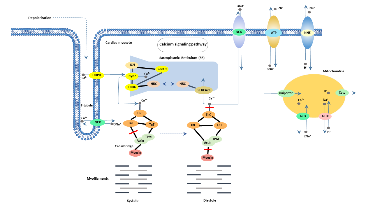
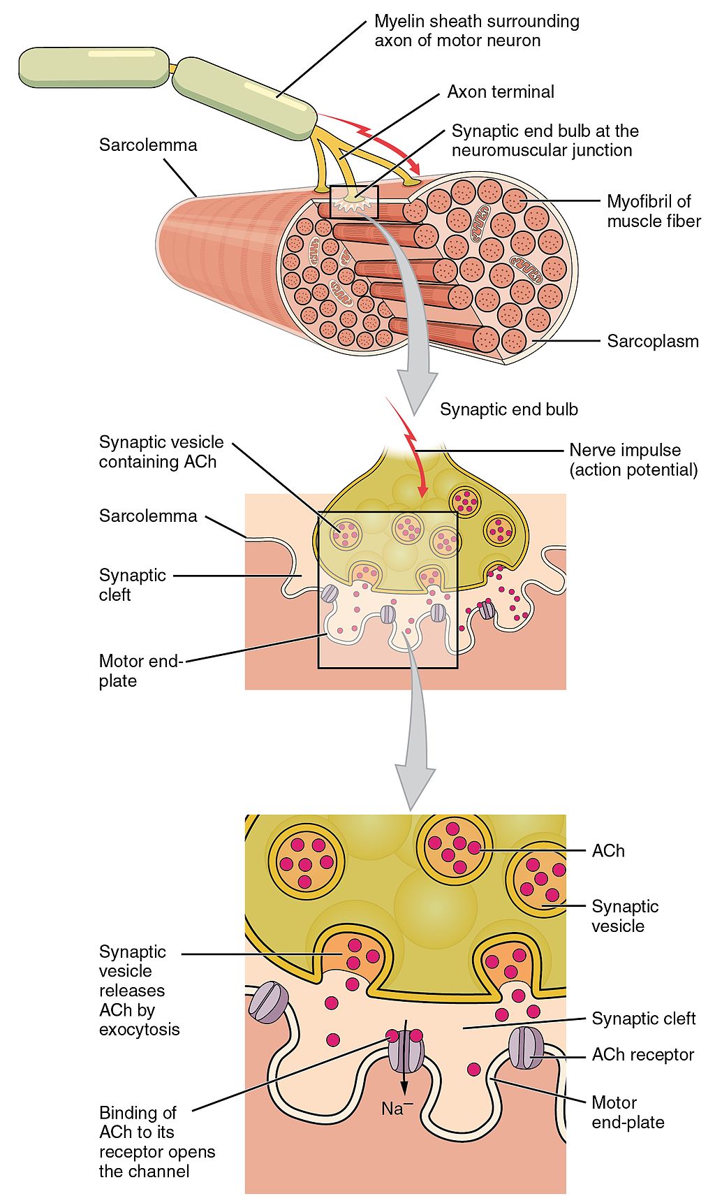
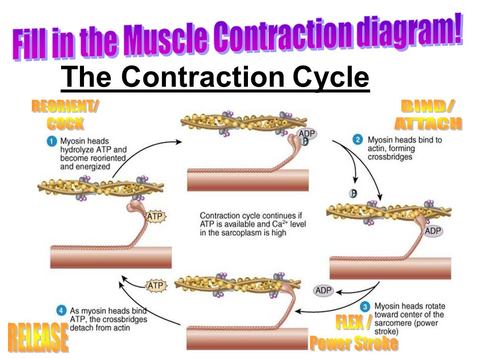
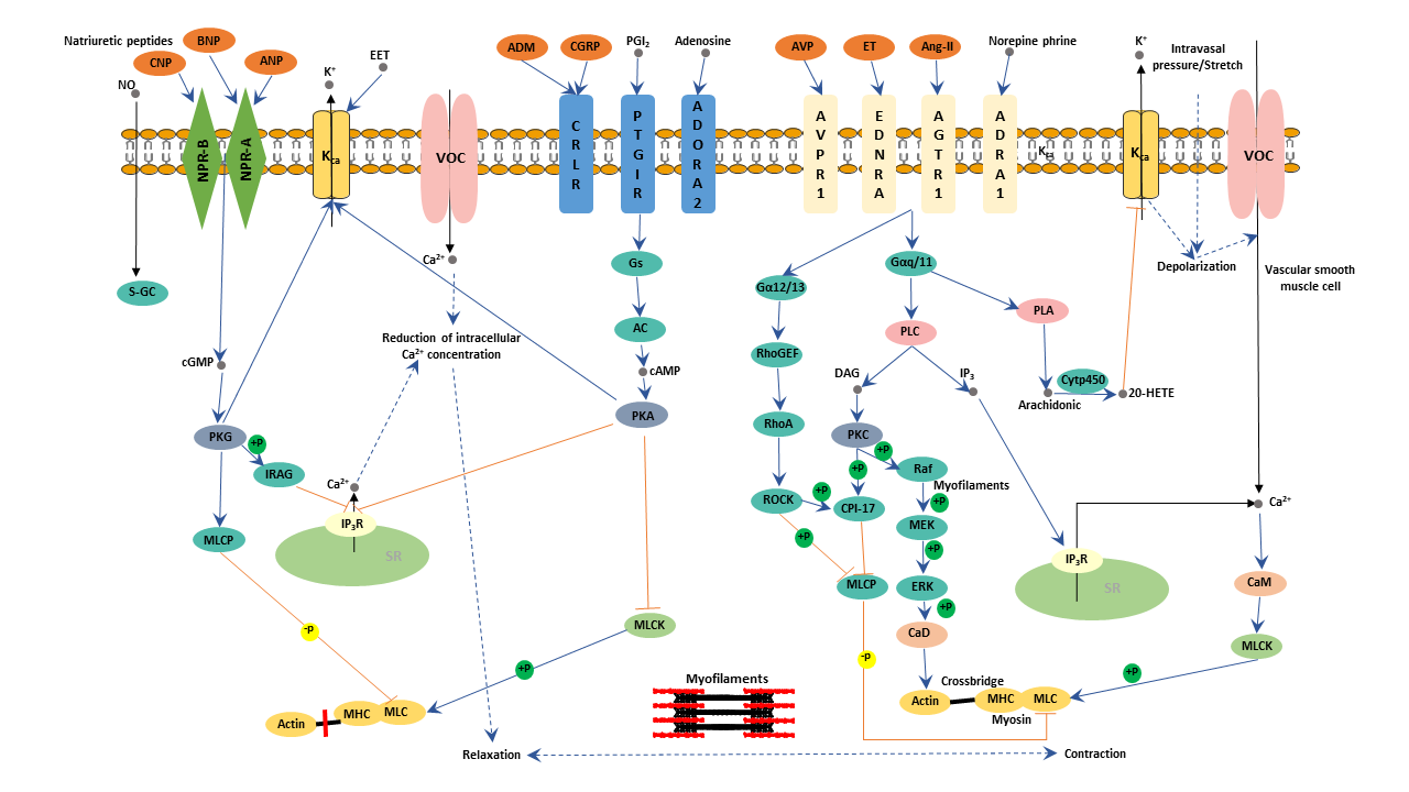
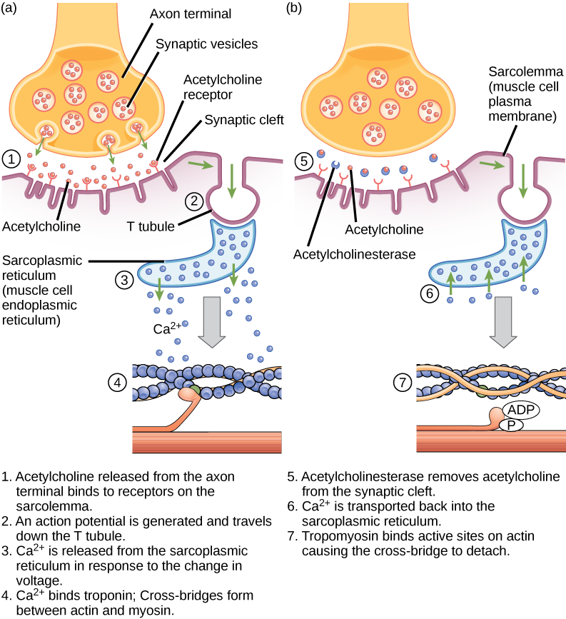
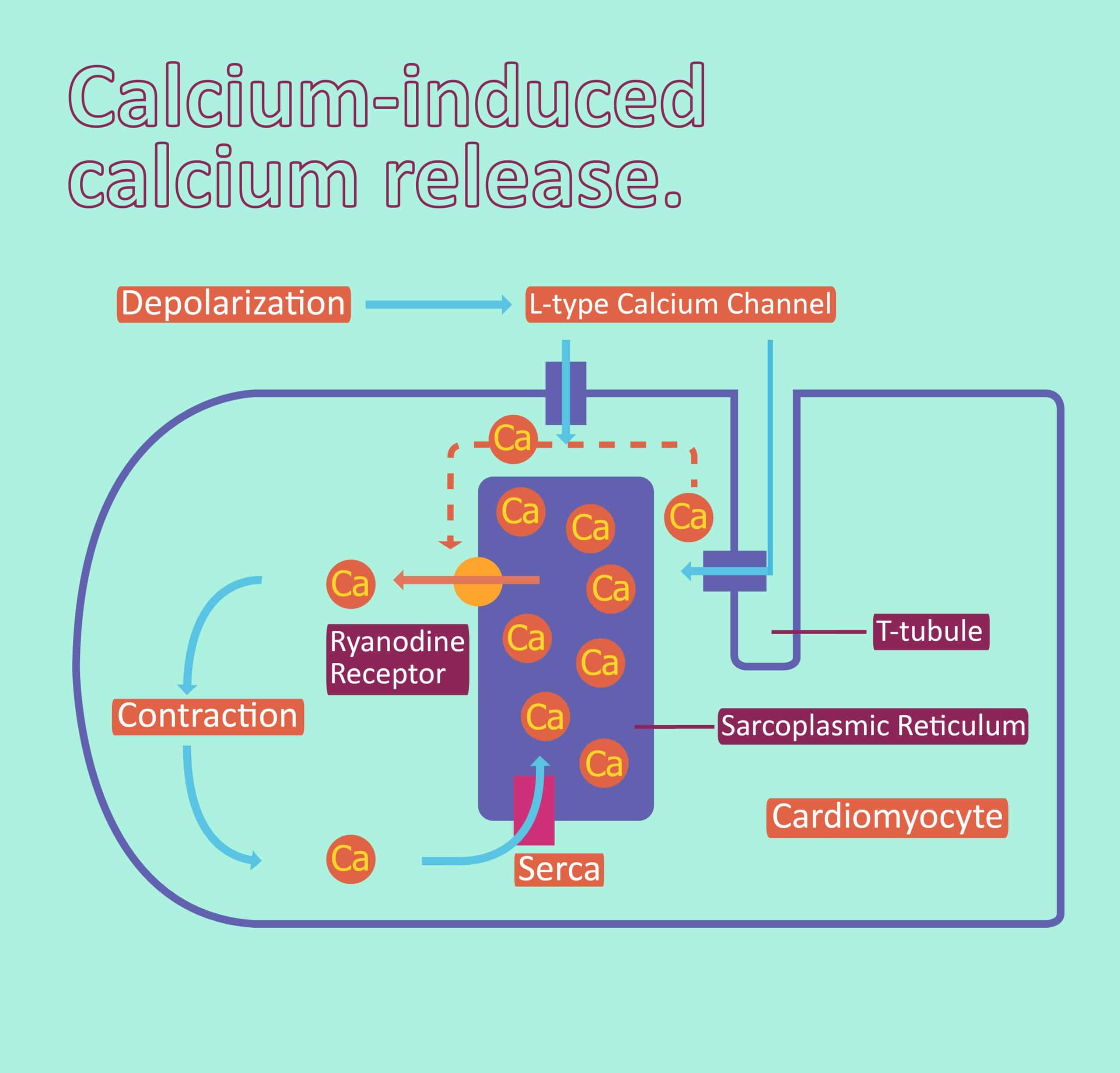
During excitation-contraction (E-C) coupling events associated with muscle force generation, Ca 2+ ions are released through the ryanodine receptor (RyR) channels in the SR membrane increasing the cytosolic [Ca 2+] to 1-2 μM for a few milliseconds.This high concentration of Ca 2+ ions facilitates the interaction of calcium with troponin to trigger the sequence of events leading to force ...

Diagram of muscle contraction
This diagram depicts muscle of the body diagrams 7441054 with parts and labels. Sarcomere - Muscle Contraction. Muscular System Muscle Diagram Muscular System Labeled Muscle Charts of the Human Body For your reference value these charts show the major superficial and deep muscles of the human body. Diagram of muscle system. There are several parts […] Muscle fibers differ in contraction speed (i.e., slow-twitch, fast-twitch) and whether they primarily generate energy aerobically or anaerobically via the glycogen-lactate system. Some of these muscles are prime movers of the arm. Gross anatomy of a skeletal muscle. Types of muscles, their structure, mechanism of their contraction and. Diagram of a nerve cell. Tête / face / dents / langue. Skeletal System from www.practitionersupplies.com.au Diagram of a nerve cell. There are around 650 skeletal muscles within the ...
Diagram of muscle contraction. Direction Of Muscle Fiber - The Muscular System /. By Drawing Maier on Kamis, 11 November 2021. Muscle contractions occur when two proteins that make up muscle fibers are activated by a nerve to increase the tension within the muscle. Caiaimage / justin pumfrey / getty images muscle contraction occurs when a muscle fiber or group of f. Actin is a globular contractile protein that interacts with myosin for muscle contraction. Skeletal muscle structure composed of muscle cells (fibers), connective tissue, blood vessels, nerves fibers are skeletal muscle tissue diagram. Actin is a globular contractile protein that interacts with myosin for muscle contraction. Each of these tissue groups is . In the body, there are three types of muscle: The three main types of muscle include skeletal, smooth and cardiac. Smooth muscle histology and diagram (inlet). Smooth muscle cells are responsible for involuntary movement, like that of the intestines during peristalsis (contraction to . (A) With minimal effort of muscle contraction, a single motor unit is seen firing at 6 Hz. The time between 2 discharges is approximately 166 milliseconds (ms), corresponding to a firing rate of 6 ...
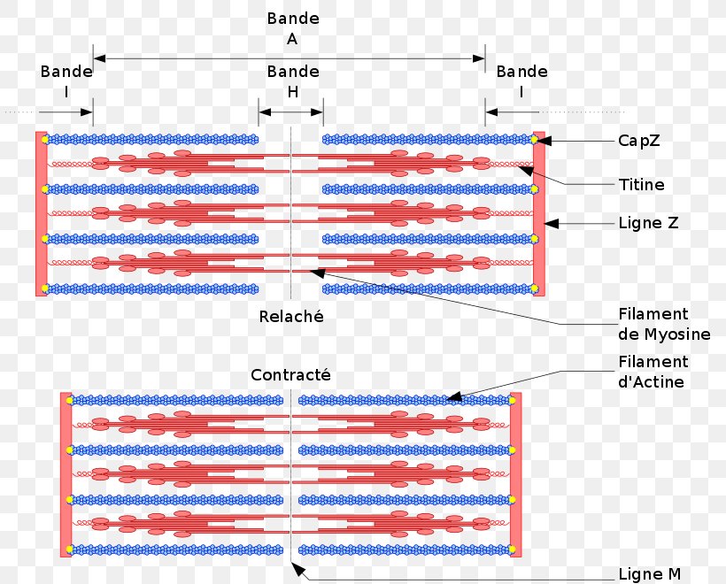
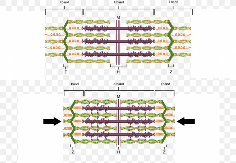
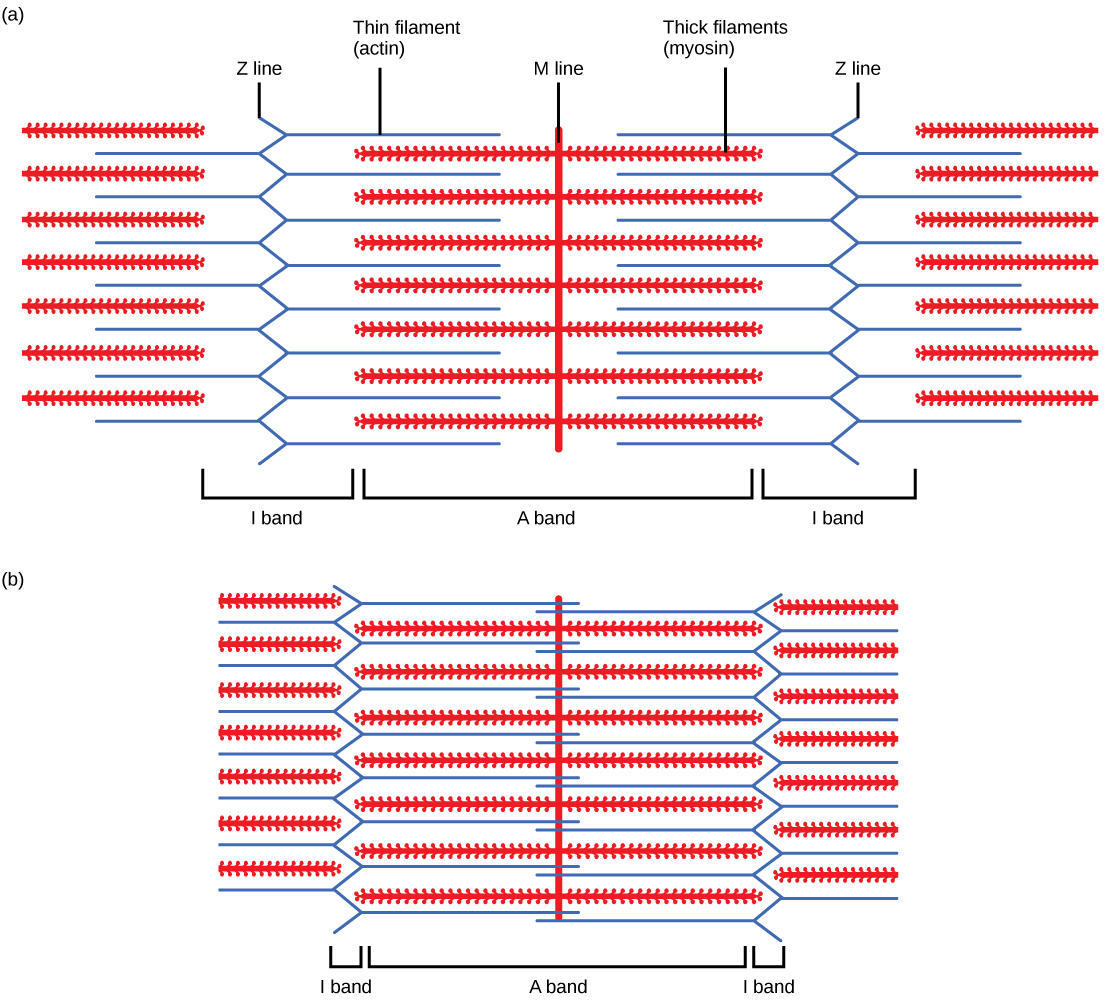
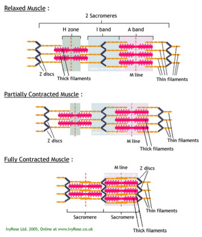
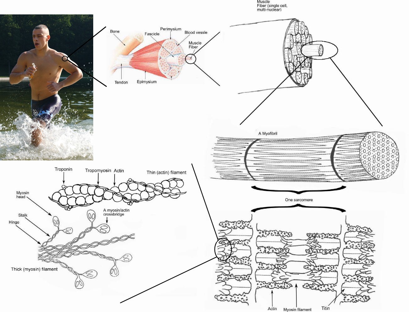
Skeletal muscles can thus produce graded contractions, the strength of which depends on the number of fibers stimulated rather than on the strength of the contractions of individual muscle fibers. If the stimulator is set to deliver an increasing frequency of electric shocks automatically, the relaxation time between successive twitches will ... 79 chase lane january 12 2021 Name_ Date_ Chapter 5 Lab Investigation. Learn vocabulary terms and more with flashcards games and other study tools. Anatomy Chapter 9 Muscular System Flashcards Quizlet Content on the quiz is the function of muscle tissue anatomy of muscle cell.Chapter 5 lab investigation muscles answer key quizlet. Chapter 5 The […] A blank diagram is a great hands on method you can use to learn the muscular system and you can also color each muscle you label and if you decide to color the muscle and its label your reinforcing the spelling of the muscle and their location in your head. Muscles are grouped together in pairs on your skeleton. Tendons are cords made of dense connective tissue and behave like bridges between muscles and bones. When a muscle contracts, the tendon moves and helps the attached bone move as well. Within the muscle fibers lies the basic functional unit of the fiber, called the sarcomere. This is where all the contraction takes place.
This diagram depicts muscle labeled diagram . Quizzes on the anatomy of the human muscular system, including the locations and actions of all the main muscles of the head and neck, the torso, and the . List the major sarcomeric proteins involved with contraction; Without muscle, humans could not live. The cardiac conduction system is a network of specialized cardiac muscle cells that initiate and transmit the electrical impulses responsible for the coordinated contractions of each cardiac cycle.These special cells are able to generate an action potential on their own (self-excitation) and pass it on to other nearby cells (conduction), including cardiomyocytes. Are called smooth muscles (nonstriated muscle). Smooth muscle histology and diagram (inlet). Smooth muscle cells are responsible for involuntary movement, like that of the intestines during peristalsis (contraction to . Cardiac muscle cells are located in the walls of the heart, appear striped (striated), and are under involuntary control. 3. Action on Smooth Muscles. With the exception of vascular muscles, acetylcholine contracts smooth muscles, and atropine has an antispasmodic response by inhibiting this acetylcholine effect. Atropine also acts in the digestive tract by decreasing amplitude, tone, and frequency of contractions.
These muscles are also known as the extrinsic eye muscles, distinguishing them from intrinsic eye muscles which are responsible for controlling the movement of It is useful to classify the extraocular muscles into two sub-groups; muscles that move the eye and muscles that move the upper eye lid. Schematic diagram of the human eye.It shows a horizontal section through the right eye.
The primary job of muscle is to move the bones of the skeleton, but muscles also enable the heart to beat and . Gross anatomy of a skeletal muscle. Muscle Diagram Labeled : Labeled Muscle Diagram Graph Diagram -. List the major sarcomeric proteins involved with contraction; Most skeletal muscles are attached to two bones through tendons.
Muscles are active in blood circulation; it is the contraction of skeletal muscles that pushes deoxygenated blood through veins and back to the heart and lungs. Muscles are also good generators of ...

Actin is a family of globular multi-functional proteins that form microfilaments in the cytoskeleton, and the thin filaments in muscle fibrils.It is found in essentially all eukaryotic cells, where it may be present at a concentration of over 100 μM; its mass is roughly 42 kDa, with a diameter of 4 to 7 nm.. An actin protein is the monomeric subunit of two types of filaments in cells ...

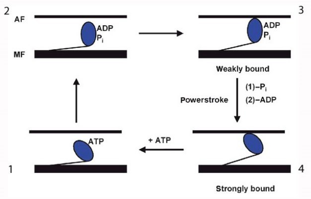
Skeletal Muscle Contraction. Skeletal muscle is striated muscle containing organized contractile structures known as sarcomeres that are made up of overlapping myofilaments: actin and myosin. When a nerve impulse arrives from a motor neuron, the signal triggers an action potential (AP) in the sarcolemma (muscle cell membrane), resulting in the ...

When an impulse travels between this space, muscle contraction happens. There are around 100-500 trillion such connections in the human brain, between two nerves or nerves and glands. In this article we will discuss only the junction. Structure of Neuromuscular Junction. As we have read, the junction consists of a neuron and a skeletal muscle cell.
During eccentric muscle contraction, the muscle _____. A. Gets longer as it contracts B. Gets shorter as it contracts C. Moves isometrically D. ... In the above diagram of an animal cell, what is the function of organelle 4? A. to store ions, create and; When there is no acceptable alternative and you cannot win, use the _____ style of conflict ...

Motor neuron and muscle contraction motor neuron the definitive guide kondo anatomy chapter 9 skeletal motor neurons in muscle contraction. Muscle Stimulation By Motor Neuron And Contraction You ... Coordination Of Independent Muscle Effectors A Conjoint Contractions Scientific Diagram

Pilates is an effective exercise method for rehabilitating musculoskeletal disorders as its principles are based on the activation of local muscles. This study aimed to compare the subjects with and without Pilates experience to find out the effect of the experience on the core muscle activity and muscle co-contraction, and to examine the relationship between the core muscle activation level ...
Vessel 53 A vasospasm is an involuntary contraction of a blood _____. First identify the structure by choosing the appropriate term from Column B and placing the corresponding answer in the answer blank. Chapter 13 - Anatomy of the Nervous System. ... Mastering A P Chapter 9 Muscle And Muscle Tissue Diagram Quizlet .
C Smooth muscle cannot stretch as much as skeletal muscle. Ch 10 3 Contraction And Relaxation Of Skeletal Muscle Fibers Flashcards Quizlet . Ch 12 13 Flashcards Quizlet . Phys Chapter 12 Muscles Flashcards Quizlet . Ch 9 Mastering A P Diagram Quizlet . Steps Of Skeletal Muscle Contraction Excitation At The Neuromuscular Junction Quiz 5 Diagram ...

Some of these muscles are prime movers of the arm. Gross anatomy of a skeletal muscle. Types of muscles, their structure, mechanism of their contraction and. Diagram of a nerve cell. Tête / face / dents / langue. Skeletal System from www.practitionersupplies.com.au Diagram of a nerve cell. There are around 650 skeletal muscles within the ...

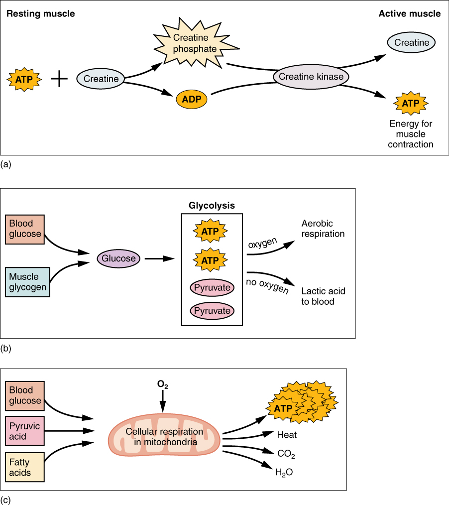
Muscle fibers differ in contraction speed (i.e., slow-twitch, fast-twitch) and whether they primarily generate energy aerobically or anaerobically via the glycogen-lactate system.
This diagram depicts muscle of the body diagrams 7441054 with parts and labels. Sarcomere - Muscle Contraction. Muscular System Muscle Diagram Muscular System Labeled Muscle Charts of the Human Body For your reference value these charts show the major superficial and deep muscles of the human body. Diagram of muscle system. There are several parts […]


0 Response to "39 diagram of muscle contraction"
Post a Comment