38 sheep brain diagram labeled
Start studying Sheep Brain Dissection labeled 2. Learn vocabulary, terms, and more with flashcards, games, and other study tools. DISSECTION OF THE SHEEP'S BRAIN Introduction The purpose of the sheep brain dissection is to familiarize you with the three-dimensional structure of the brain and teach you one of the great methods of studying the brain: looking at its structure. One of the great truths of studying biology is the saying that "anatomy precedes physiology".
Sheep Brain Anatomy #2. STUDY. PLAY. cerebellum. posterior part of the brain that coordinates muscle movements and maintains balance. temporal lobe. interpretation and integration of speech and sound. parietal lobe. interpretation and integration of sensory stimuli. frontal lobe.
Sheep brain diagram labeled
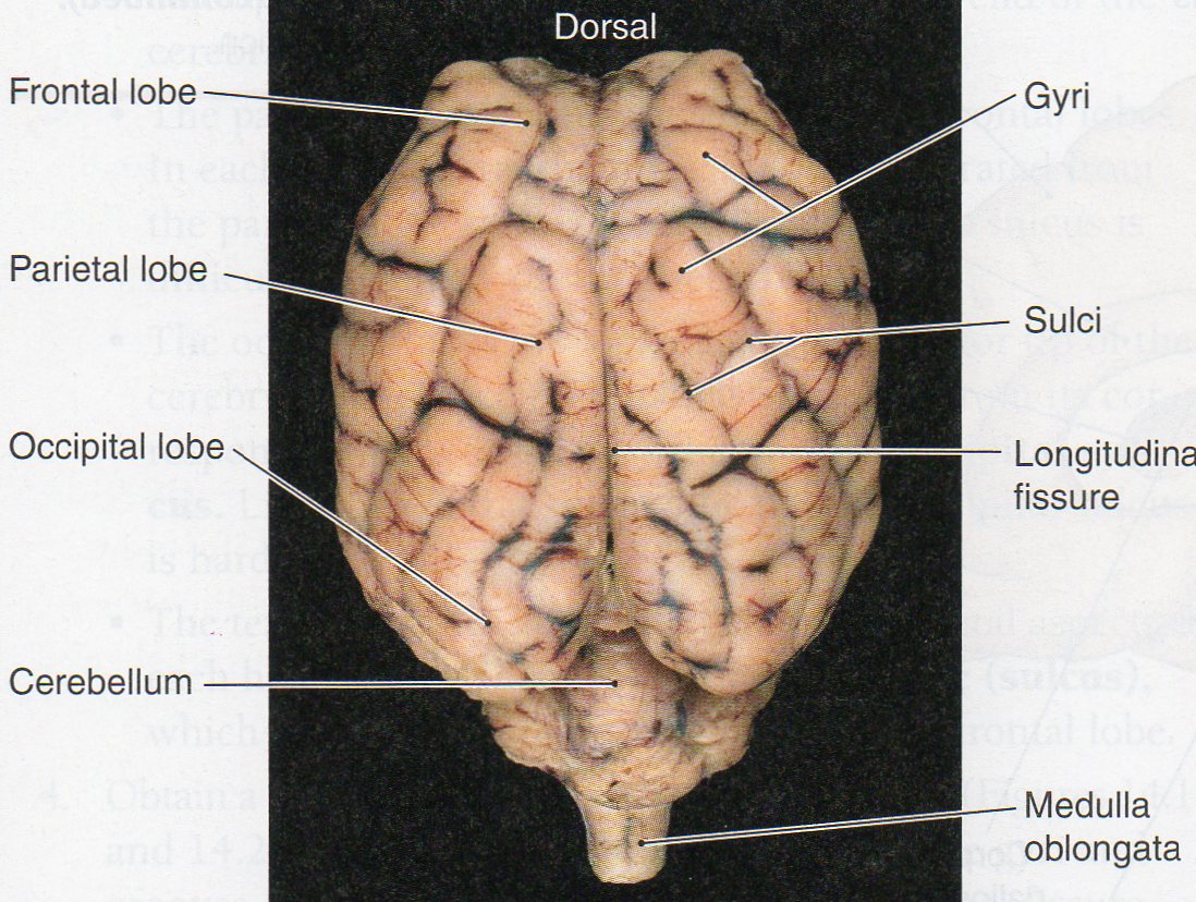
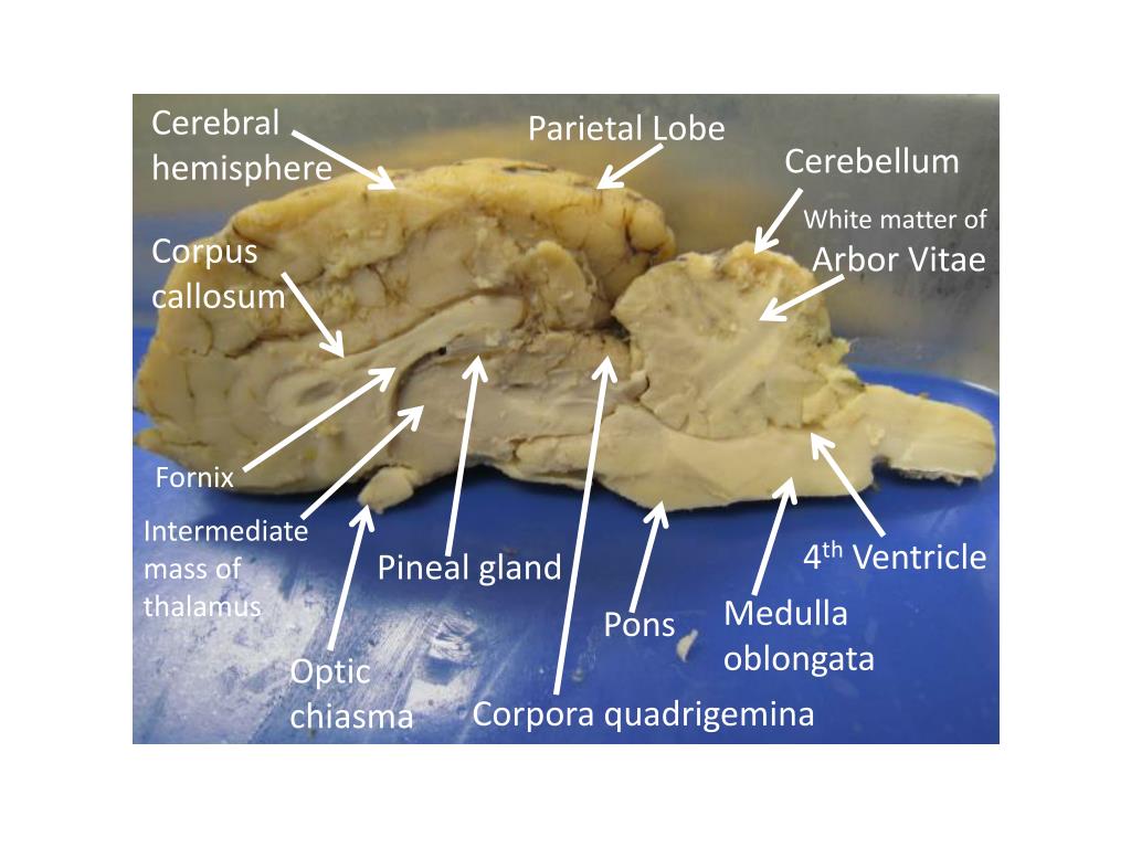
The sheep brain is exposed and each of the structures are labeled and described in a sequential manner, in the same way that a real dissection would occur. Amanda Huss Anatomy Sheep Neuroanatomy Lab- Labeling Worksheet Psychology 2315- Brain and Behaviour Kwantlen Polytechnic University Figure 1: Dorsal view Cerebellum, Frontal lobe, Occipital lobe, Parietal lobe, and Temporal lobe. Temporal Parietal Lobe Frontal Lobe Cerebellum Occipital Lobe Diagram Worksheets. Label the Parts of a Sheep Brain. Print out these diagrams and fill in the labels to test your knowledge of sheep brain anatomy. Internal anatomy: label the right side (.pdf) External anatomy: label the top view (.pdf) External anatomy: label the bottom view (.pdf) What other users say: Fun and Educational.
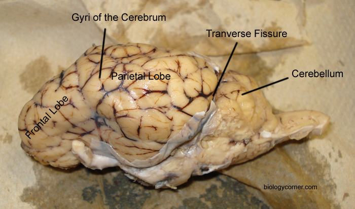
Sheep brain diagram labeled. Diagram of Sheep Brain - Lateral view Label theBrain of the Sheep. Publisher: Biologycorner.com; follow on Google+ This work is licensed under a Creative Commons Attribution-NonCommercial 3.0 Unported License. Brain Label Answer Key. Image adapted from a photograph of the sheep brain. ... Sheep Brain Coronal Cut D. 18 terms. kph43. Sheep Brain Coronal Cut C. 15 terms. kph43. Upgrade to remove ads. Only $2.99/month. labeled brain Brain Anatomy, Human Anatomy And Physiology, Medical Anatomy, ... Images taken from the dissection of the sheep's brain: cerebrum, cerebellum, ...
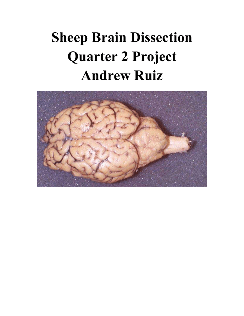
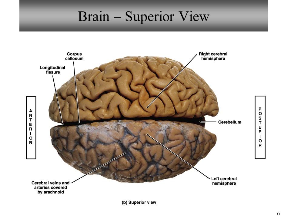
The sheep brain is exposed and each of the structures are labeled and described in a sequential manner, in the same way that a real dissection would occur. Sheep Brain Dissection. 1. The sheep brain is enclosed in a tough outer covering called the dura mater. You can still see some structures on the brain before you remove the dura mater. The sheep brain is quite similar to the human brain except for proportion. The sheep has a smaller cerebrum. Also, the sheep brain is oriented anterior to ... The lobes of the brain are visible, as well as the transverse fissure, which separates the cerebrum from the cerebellum. The convolutions of the brain are also visible as bumps (gyri) and grooves (sulci). Use the diagram below to help you locate these items. Dorsal View of the Sheep Brain . 8. Image Result For Sheep Brain Labeled Brain Diagram Human Brain Diagram Brain Anatomy. Sheep Brain Dissection Project Guide Hst Learning Center Dissection Brain Mapping Science Biology. Sheep Brain Dissection Lab Companion In 2021 Brain Anatomy Anatomy And Physiology Brain. Sheep Brain External View Labeled Anatomia Veterinaria Anatomia Veterinaria.
Dissection Instructions: Obtain a preserved sheep brain from the bucket in the front of the classroom. Place this on your dissection tray. You will need the following dissection tools to properly perform this lab: scalpel. scissors. probes. 3. The sheep brain is enclosed in a tough outer covering called the dura mater. Sheep Brain Anatomy Lab Manual. Based on original material by R. N. Leaton, Dartmouth College. Contributors to this version: Al Sorenson, Lisa Raskin, Sarah Turgeon, Steve George, and JP Baird. I. Introduction. The brain of the sheep is useful for study because its anatomy is similar to human brain anatomy. Although exact proportions (and names ... function, and pathology. Those students participating in Sheep Brain Dissections will have the opportunity to dissect and compare anatomical structures. At the end of this document, you will find anatomical diagrams, vocabulary review, and pre/post tests for your students. The following topics will be covered: 1. to anatomy studies. See for yourself what the . cerebrum, cerebellum, spinal cord, gray matter, white matter, and other parts of the brain look like! Observation: External Anatomy . 1. You'll need a . preserved sheep brain. for the dissection. Set the brain down so the flatter side, with the white . spinal cord. at one end, rests on the ...
Start studying Sheep Brain Dissection labeled. Learn vocabulary, terms, and more with flashcards, games, and other study tools.
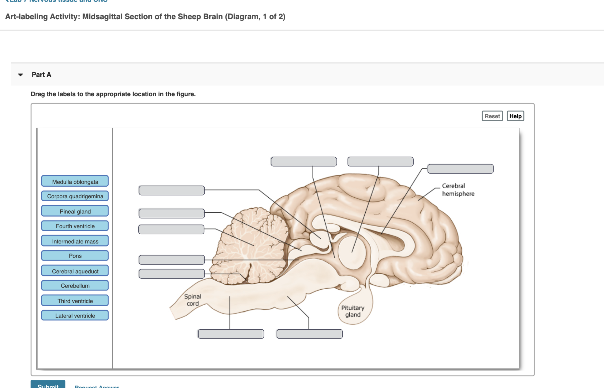
5 3 11 6 22 16 18 1. Gray Matter 2. White Matter 3. Corpus Callosum 4. Lateral Ventricle 5. Caudate Nucleus 6. Septum Pellucidum 7. Fornix 8.
NERVOUS SYSTEM - SHEEP BRAIN IMAGES. Sheep Brain Unlabeled. Sheep Brain in Dura Mater · Dorsal Sheep Brain · Dorsal-Colliculi ... Sheep Brain Labeled.
Sheep Brain Dissection with Labeled Images. The sheep brain is exposed and each of the structures are labeled and described in a sequential manner, in the same way that a real dissection would occur. amhuss. A. Amanda Huss. Anatomy. Brain Anatomy. Human Anatomy And Physiology. Medical Anatomy.
Sheep are wonderful and cute. The brain is an interesting organ. It helps with cognition and memory. Almost all the basic task In the body is commanded by the Brain. It is the control center of the body which regulates and control the process crucial for survival Are you interested in learning more about the brain of different animals? Can you answer all the questions of this "Sheep Brain ...
View now! Pretty good picture of the sheep brain labeled. Central Nervous System Worksheet coloring page from Anatomy category. Select from 36976 printable crafts of cartoons, nature, animals, Bible and many more. Shows pictures of a sheep and a human brain.
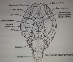
Diagram Worksheets. Label the Parts of a Sheep Brain. Print out these diagrams and fill in the labels to test your knowledge of sheep brain anatomy. Internal anatomy: label the right side (.pdf) External anatomy: label the top view (.pdf) External anatomy: label the bottom view (.pdf) What other users say: Fun and Educational.
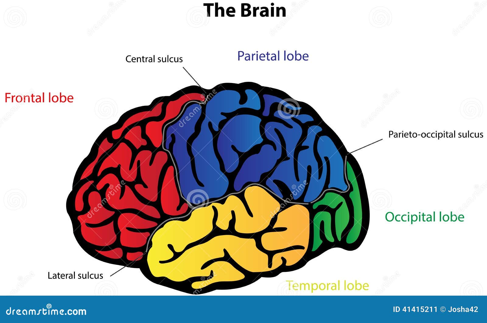
Sheep Neuroanatomy Lab- Labeling Worksheet Psychology 2315- Brain and Behaviour Kwantlen Polytechnic University Figure 1: Dorsal view Cerebellum, Frontal lobe, Occipital lobe, Parietal lobe, and Temporal lobe. Temporal Parietal Lobe Frontal Lobe Cerebellum Occipital Lobe
The sheep brain is exposed and each of the structures are labeled and described in a sequential manner, in the same way that a real dissection would occur. Amanda Huss Anatomy










(294).jpg)

0 Response to "38 sheep brain diagram labeled"
Post a Comment