38 diagram of synovial joints
Carpal Bone Joints. All the joints involving the carpal bones are synovial joints, where the articulation surface has a flexible cartilage layer, along with a fluid lining to allow for better freedom of movement [22].. The Radiocarpal Joint: Those between the radius and the proximal carpal bones (except pisiform) [8]. Intercarpal Joints: Articulations between the carpal bones in hand are an ... Find synovial joints stock images in HD and millions of other royalty-free stock photos, illustrations and vectors in the ... Synovial joint diagram.
08-11-2019 · Hinge joints allow bones to move in one direction back and forth, much like the hinge on a door. This article looks at their anatomy and function and includes an interactive diagram.

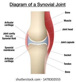
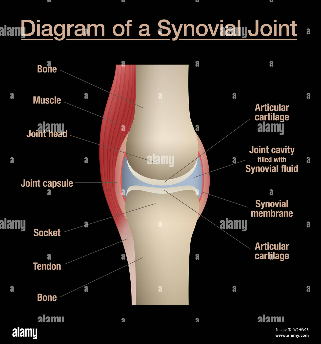
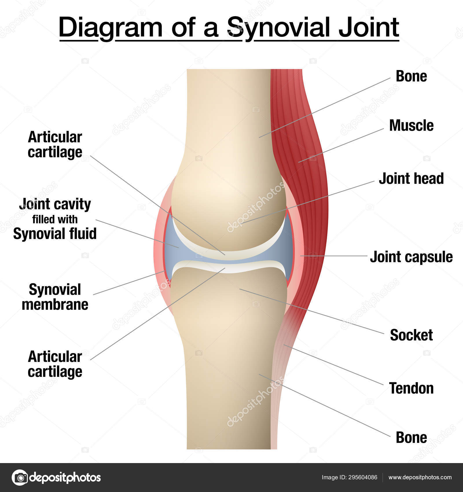
Diagram of synovial joints
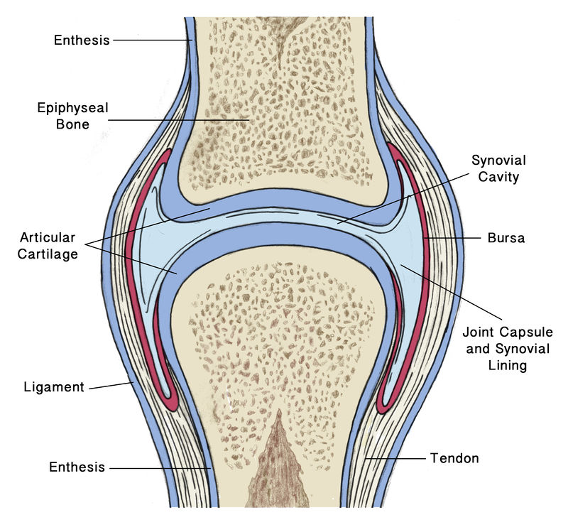
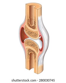
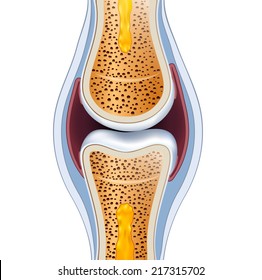
Download scientific diagram | A general synovial joint. from publication: Homeostasis 6: Nurses as external control agents in rheumatoid arthritis | All ... 05-11-2021 · Synovial joints, the most movable joints in our bodies, allow us to perform actions such as bending our arms at the elbow. Learn to define the six types of … A synovial joint is characterised by the presence of a fluid-filled joint cavity contained within a fibrous capsule. It is the most common type of joint ...
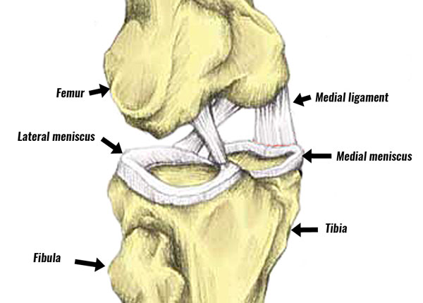
Diagram of synovial joints. A synovial joint or diarthrosis occurs at articulating bones to allow movement. ... Elbow Joint: Diagram of the anastomosis around the elbow joint. 20-03-2021 · Hero Images / Getty Images. The location of the knee pain can be useful information when trying to obtain an accurate diagnosis. Pain at the front of the knee can be caused by bursitis, arthritis, or softening of the patella cartilage, as in chrondromalacia patella.. Pain on the side of the knee is usually associated with injury to the collateral ligaments, arthritis, or tears to the menisci. Synovial joints are often further classified by the type of movements they permit. There are six such classifications: hinge (elbow), saddle (carpometacarpal ... Nov 3, 2013 - synovial joint. ... Diagram of a synovial joint ... increase bone cartilage Synovial Joint, Bursitis Hip, Body Joints, Rheumatoid Arthritis, ...
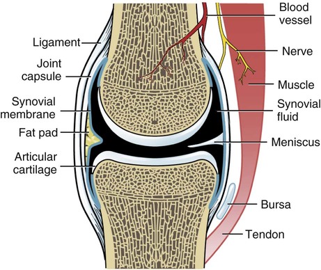
A joint or articulation (or articular surface) is the connection made between bones in the body which link the skeletal system into a functional whole. They are constructed to allow for different degrees and types of movement. Some joints, such as the knee, elbow, and shoulder, are self-lubricating, almost frictionless, and are able to withstand compression and maintain heavy loads while still ... 01-01-2019 · The synovial fluid is similar to raw egg whites in its consistency. The function of the bursae and the synovial fluid is to reduce the friction that occurs as ligament and bones, ligaments and tendons, or tendons and bones rub together. Joints have bursae around them, and because the hip joint is so large it has many bursae, around 20 total. These joints form the most prominent knuckles of the hand [7]. Carpometacarpal Joints (Carpal-Metacarpal Joints) The joints between the metacarpal and carpal bones, these are all plane synovial joints, except the thumb as it is a saddle joint (another form of synovial joint) [8]. Types of synovial joints. There are six types of synovial joints which allow varying types and ranges of movement to occur. The variation in the movements at these joints is because of the differences in their characteristics and limiting factors, as previously discussed. The six synovial joints are: 1.
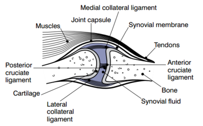
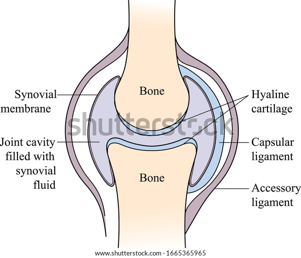
Note: Synovial joint joins bones or cartilage with a fibrous joint capsule which continues with the periosteum of the bone, and forms the outer boundary of a ...1 answer · Top answer: Hint: A synovial joint is the type of joint found between two bones that move against each other, such as the joints of the limbs (e.g. shoulder, hip, elbow ... Types of Synovial Joints. Synovial joints are subdivided based on the shapes of the articulating surfaces of the bones that form each joint. The six types of synovial joints are pivot, hinge, condyloid, saddle, plane, and ball-and socket-joints (Figure 9.4.3).Figure 9.4.3 – Types of Synovial Joints: The six types of synovial joints allow the body to move in a variety of ways. The synovial cavity/joint is filled with synovial fluid. The joint capsule is made up of an outer layer, the articular capsule, which keeps the bones together ... 07-09-2021 · Dog leg anatomy joints. You know, joints are formed when two or more bones are joined by fibrous, elastic, or cartilaginous tissues or by a combination of these. I have a detailed article on the joint anatomy of an animal here. Ensure you know the anatomy of different types of joints like fibrous, cartilaginous, and synovial joints of an animal ...
A synovial joint is characterised by the presence of a fluid-filled joint cavity contained within a fibrous capsule. It is the most common type of joint ...
05-11-2021 · Synovial joints, the most movable joints in our bodies, allow us to perform actions such as bending our arms at the elbow. Learn to define the six types of …
Download scientific diagram | A general synovial joint. from publication: Homeostasis 6: Nurses as external control agents in rheumatoid arthritis | All ...


:max_bytes(150000):strip_icc()/types_of_synovial_joints-5b6f001e4cedfd00251806ae.jpg)


0 Response to "38 diagram of synovial joints"
Post a Comment