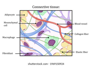
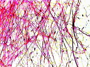
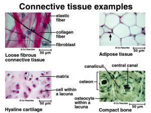
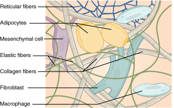
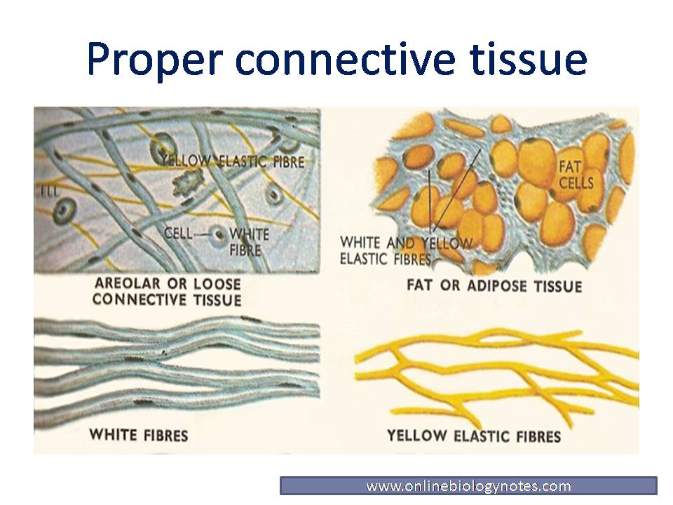
38 areolar connective tissue diagram
Can yall answer these set of questions pls. *Q1*:-Describe the following tissue with the help of neat and well labelled diagram :- 1)Squamous epitheli … al tissue 2) Cuboidal epithelial tissue 3) Glandular epithelial tissue 4) Columnar epithelial tissue 5) Ciliated epithelial tissue *Q2*:-Write a note on- Areolar Connective tissue *Q.3 ... Sep 18, 2021 · Areolar Tissue. Areolar tissue is the most widely distributed solid connective tissue in the body. Which makes sense because areolar tissue is present under the entire skin. And areolar tissue also supports the epithelium. Areolar tissue contains randomly spread out fibers, fibroblasts, mast cells, and macrophages.
the word fiber is used to describe a structure in three of the four primary (main) tissue types. In connective tissues, a fiber is a protein strand that is collagen, reticular, or elastic. The meaning of the work fiber is different in muscle tissue and in nervous tissue. Explain a fiber in each of these two tissue types.

Areolar connective tissue diagram
MCQ on Animal Tissue. Q1. Categorization of epithelial tissue is based on this. Q2. Mast cells are linked to. Q3. In connective tissue sheaths, this is the correct sequence stretching from the outermost to the innermost layer. Q4. This is not a function of neuroglia. Examination includes breast tissue bounded by the clavicle, sternal border, in ramammary crease, and midaxillary line. Breast palpation within this pentagonal area is approached in a linear ashion. echnique uses the nger pads in a continuous rolling, gliding circular motion (Fig. 1-4). Learn how areolar connective tissue differs from other tissue types. Large Intestine Anatomy, Parts, Diagram & Major Function Discuss the role of the large intestine in humans, the anatomy, and ...
Areolar connective tissue diagram. Can yall answer these set of questions pls. *Q1*:-Describe the following tissue with the help of neat and well labelled diagram :- 1)Squamous epitheli … al tissue 2) Cuboidal epithelial tissue 3) Glandular epithelial tissue 4) Columnar epithelial tissue 5) Ciliated epithelial tissue *Q2*:-Write a note on- Areolar Connective tissue *Q.3 ... A tissue however, other cellular features such as cilia may also be described in the classification system. In biology, tissue is a cellular organizational level between cells and a complete organ. A tissue is an ensemble of similar cells and their extracellular matrix from the same origin that together carry out a specific function. II. Areolar Connective Tissue - a loosely arranged fibroelastic connective tissue. Unstained, it appears translucent, soft and syncitial. A. Compostion. 1. Cells - All cell types associated with connective tissues may be seen in areolar. Fibroblasts - are the most numerous cells in areolar. The cells are large, flat and branching or spindle-shaped. Cartilage is a robust and viscoelastic connective tissue that can be found in joints between bones, the rib cage, intervertebral discs, the ear, and the nose. While more rigid and less flexible than muscle, cartilage is not as stiff as bone. These properties allow cartilage to serve as a support structure for holding tubes open or for .
Nov 05, 2021 · Loose Connective Tissue . It comprises areolar tissue, adipose tissue, tendons and ligaments. 1. Areolar tissue: This tissue binds our skin to the underlying tissues. They form the mass of our body. This is also called loose connective tissue. This is one of the most common and abundant loose connective tissues. They contain fibroblasts and ... The gastrointestinal tract (GI tract, GIT, digestive tract, digestion tract, alimentary canal) is the tract from the mouth to the anus which includes all the organs of the digestive system in humans and other animals.Food taken in through the mouth is digested to extract nutrients and absorb energy, and the waste expelled as feces.The mouth, esophagus, stomach and intestines are all part of ... Female reproductive system diagram labeled. Learn vocabulary terms and more with flashcards games and other study tools. These human anatomy models are of the highest quality and include such pieces as male reproductive system anatomy model and female reproductive system anatomy model anatomical fetus models in various stages of growth birthing. Apr 05, 2021 · i. Areolar connective tissue. Areolar tissue is one of the most widely distributed connective tissues commonly found in nearly each body structure. This tissue consists of cells, fibers, and ground substances in roughly equal parts. The most abundant cells are fibroblasts, but other types of cells are also found in limited concentration.
Diagram of a neuron along with the labelling is as follows: 10. Name the following. (a) Tissue that forms the inner lining of our mouth. (b) Tissue that connects muscle to bone in humans. (c) Tissue that transports food in plants. (d) Tissue that stores fat in our body. (e) Connective tissue with a fluid matrix. (f) Tissue present in the brain ... May 14, 2021 · Areolar Connective Tissue Diagram. This diagram shows areolar connective tissue serving as a medium between epithelial tissue and blood vessels. Cellular components (such as fibroblasts) and the ... Connective tissue flow chart ncert. This tissue is present in the skin. Further there are. Areolar connective tissue. Areolar tissue fills the space inside the organs. Show the types of animal tissues using flow chart. A Dense regular b Dense irregular. Examples include blood bones cartilages tendons ligaments areolar tissues and adipose tissues. May 09, 2021 · Areolar connective tissue – The areolar connective tissue is a loose array of fibers consists of various types of cells. It is usually located under the epithelia; which is the outer covering of the blood vessel including the esophagus, fascia between muscles, pericardial sacs, and other organs of the body.
Learn how areolar connective tissue differs from other tissue types. Large Intestine Anatomy, Parts, Diagram & Major Function Discuss the role of the large intestine in humans, the anatomy, and ...
Examination includes breast tissue bounded by the clavicle, sternal border, in ramammary crease, and midaxillary line. Breast palpation within this pentagonal area is approached in a linear ashion. echnique uses the nger pads in a continuous rolling, gliding circular motion (Fig. 1-4).
MCQ on Animal Tissue. Q1. Categorization of epithelial tissue is based on this. Q2. Mast cells are linked to. Q3. In connective tissue sheaths, this is the correct sequence stretching from the outermost to the innermost layer. Q4. This is not a function of neuroglia.










0 Response to "38 areolar connective tissue diagram"
Post a Comment