39 atomic force microscopy diagram
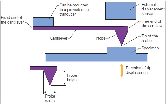
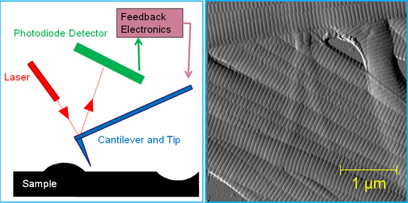
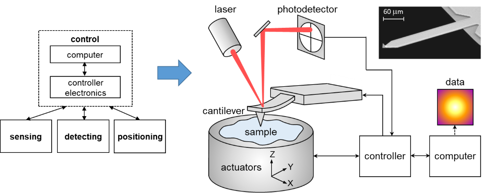
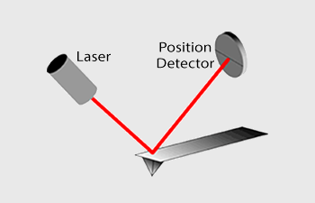
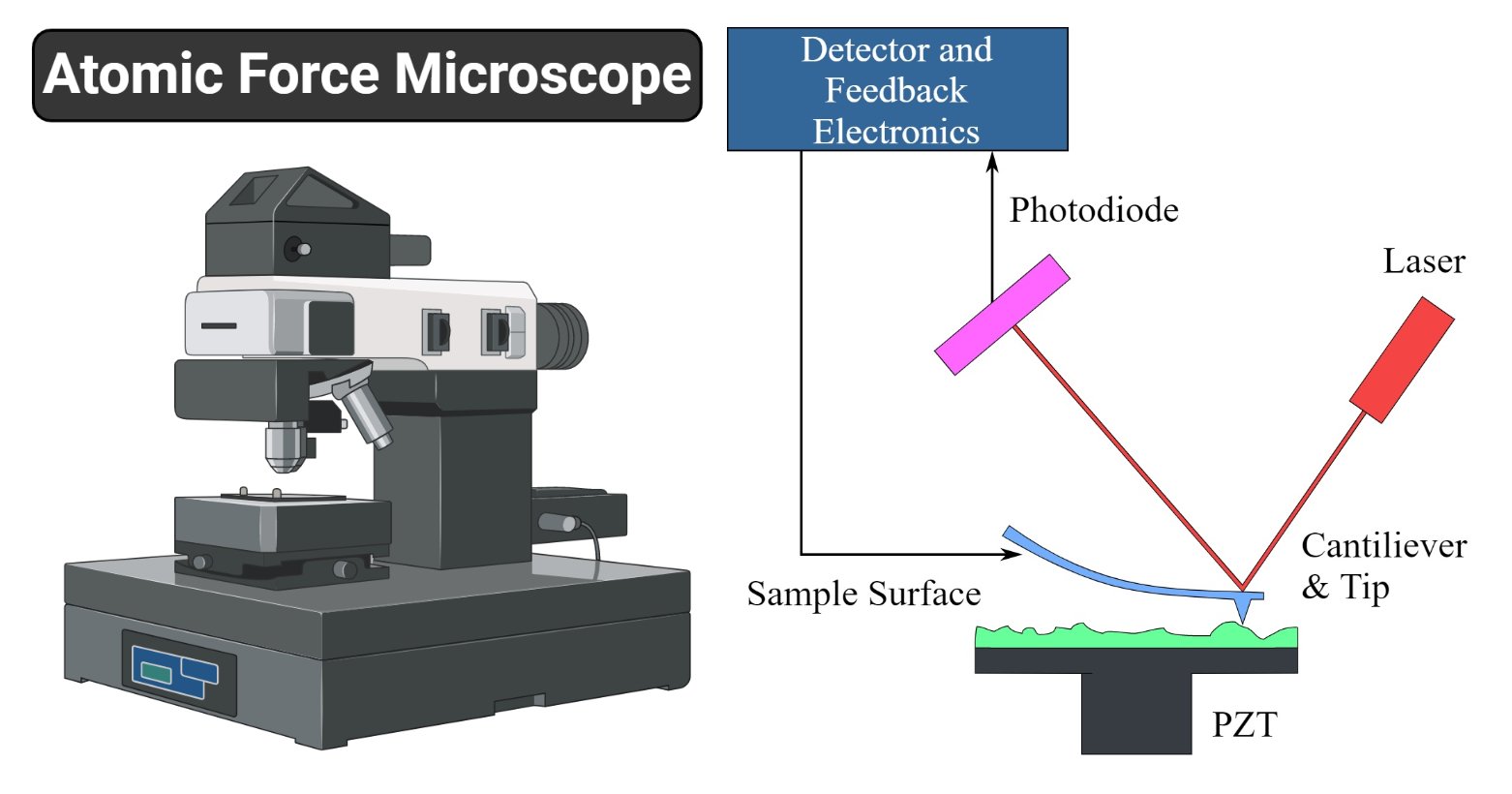
The AFM principle is based on the cantilever/tip assembly that interacts with the sample; this assembly is also commonly referred to as the probe. The AFM probe interacts with the substrate through a raster scanning motion. The up/down and side to side motion of the AFM tip as it scans along the surface is monitored through a laser beam reflected off the cantilever. Park Systems, a world leading manufacturer of Atomic Force Microscopes, presents Park NX-Hybrid WLI, the first fully integrated system that combines Atomic Force Microscopy (AFM) with White Light ...
Atomic-force microscopy is a reference method for traceable and correlative measurements of nanostructures. The Nanostructure Fabrication and Measurement Group is developing critical-dimension and traceable microscope systems to calibrate probe tips and microscopy standards, and measure diverse devices ranging from waveguides to nanoparticles.

Atomic force microscopy diagram
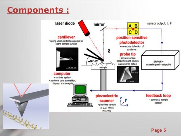
In this article we will discuss about the design of atomic force microscope, explained with the help of a diagram. An atomic force microscope instead of using a lens is provided with a probe to examine the surface of a specimen with a sharp tip which may be several micrometers in and less than 10 nm in diameter at the point near field to be examined. The tip lies at the end of the lever which ... Scanning probe microscopy has been the engine of characterization in nanoscale systems ().Atomic force microscopy (AFM) in particular has developed into a leading technique for high-resolution studies without material restrictions (3-5).It is increasingly being used for detailed characterization in a wide variety of physical, biological, and chemical processes (6, 7). Functionalizing the tip of an atomic force microscope with a CO molecule enabled atomic-resolution imaging of single molecules, and measurement of their adsorption geometry and bond-order relations. In addition, by using scanning tunneling microscopy and Kelvin probe force microscopy, the density of the molecular frontier orbitals and the ...
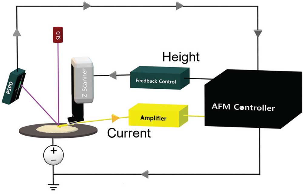
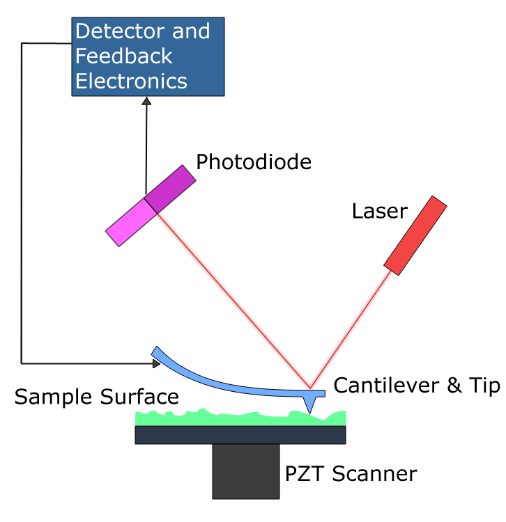
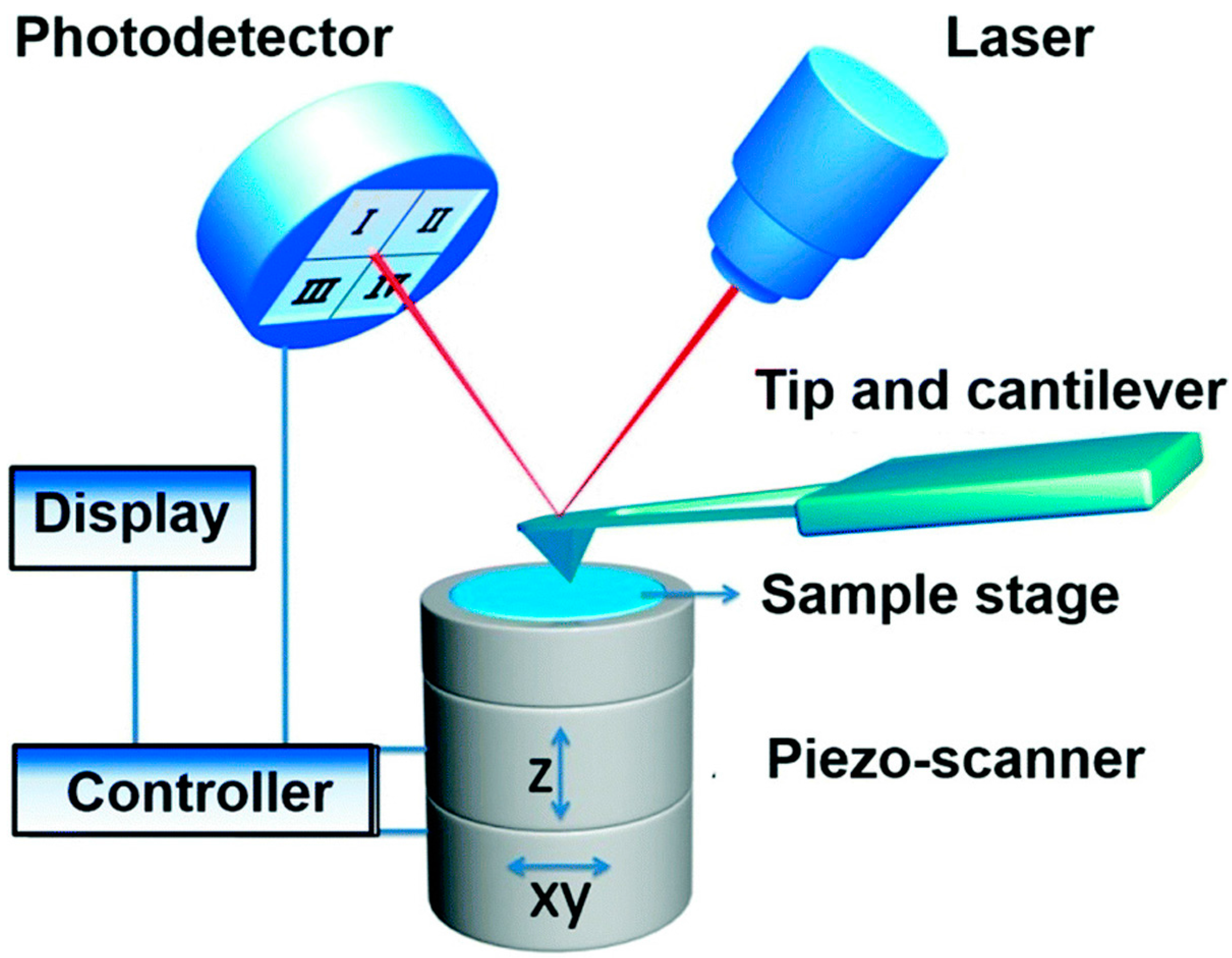
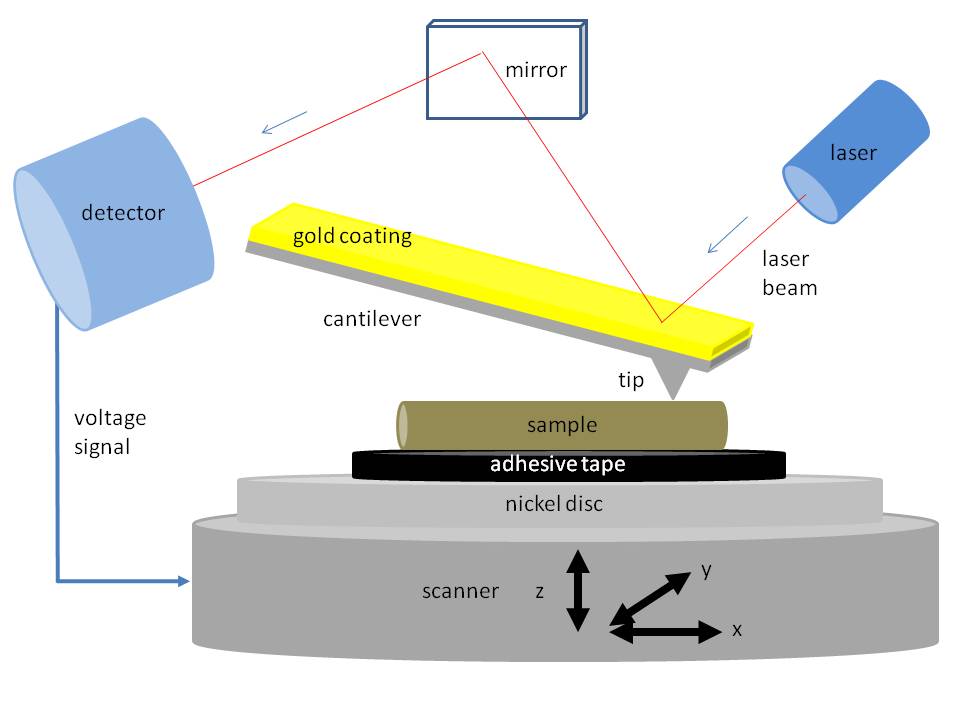
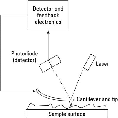
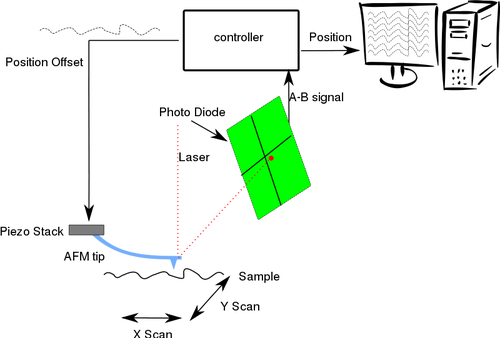
Atomic force microscopy diagram. The Atomic Force Microscope (AFM) takes the image of the surface topography of the sample by force by scanning the cantilever over a section of interest. Depending on how raised or how low the surface of the sample is, it determines the deflection of the beam, which is monitored by the Positive-sensitive photo-diode (PSDP). 2.1. Atomic Force Microscopy (AFM) AFM is a topographic imaging technique with high spatial resolution (a lateral resolution of 1 nm and a vertical resolution of 0.1 nm), and can be used to acquire mechanical properties [].The principle of AFM (Figure 1) is that the sample can be imaged at atomic resolution by detecting the near-field interactions between a tiny tip and the sample surface [17,18]. File:Atomic force microscope block diagram.svg. Size of this PNG preview of this SVG file: 646 × 599 pixels. Other resolutions: 259 × 240 pixels | 517 × 480 pixels | 647 × 600 pixels | 828 × 768 pixels | 1,104 × 1,024 pixels | 926 × 859 pixels. AFM—atomic force microscopy. Atomic force microscopy (AFM) is a kind of scanning probe microscopy, where a probe or tip is used to map the contours of the sample. During operational mode, the tip connected to a cantilever is scanned over the surface of the sample, with a small repulsive force present between the sample and the tip.
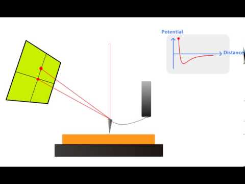
Atomic force microscopy (AFM) is a type of scanning probe microscopy (SPM), with demonstrated resolution on the order of fractions of a nanometer, more than 1000 times better than the optical diffraction limit.The information is gathered by "feeling" or "touching" the surface with a mechanical probe. Piezoelectric elements that facilitate tiny but accurate and precise movements on (electronic ... Press Release Atomic-Force Microscopy (AFM) Market 2021: Comprehensive Growth, Industry Size, Share, Emerging Trends, Revenue, COVID-19 Impact Analysis, Demand, Gross Margin and Forecast 2025 with ... Visualization of cell structure by atomic force microscopy. MOJ Anat Physiol. 2017;3(5):160‒161. DOI: 10.15406/mojap.2017.03.00109 Discussion Microscopes produce images of cell that may further be analyzed. While the light and electron microscopes use lenses and a source of light or electrons, atomic force microscopes use a very fine tip that Atomic Force Microscopy (AFM) 1. General Principle The Atomic Force Microscope is a kind of scanning probe microscope in which a topographical image of the sample surface can be achieved based on the interactions between a tip and a sample surface. The atomic force microscope was invented by Gerd Binning et al. in 1986 at IBM Zurich based on ...
Download scientific diagram | Schematic drawing of the atomic force microscope. from publication: Direct Measurement of Interaction Forces between Surfaces in Liquids Using Atomic Force Microscopy ... Atomic force microscopy (AFM) is a tool for studying the microscopic world. Capable of using and measuring van der Waals forces, AFM is a new super-resolution near-field probe microscopic technique that can be applied to both conductors and insulators. Most existing indirect or calcula-tion procedures estimate the internal surface structure of Keywords:Atomic force microscopy, Protein structure, Amyloid, Disease, Correlative microscopy, NMR. Abstract:Proteins are versatile macromolecules that perform a variety of functions and participate in virtually all cellular processes. The functionality of a protein greatly depends on its structure and alterations may result in the development ... §D. Sarid, Scanning Force Microscopy with Applications to Electric, Magnetic and Atomic Forces , Revised Edition, Oxford University Press, 1994. § U. Dürig, "Interaction sensing in dynamic force microscopy", New Journal of
An Introductory Guide To Atomic Force Microscopy Afm Tsc
Atomic force microscopy (AFM) was developed when people tried to extend STM technique to investigate the electrically non-conductive materials, like proteins. In 1986, Binnig and Quate demonstrated for the first time the ideas of AFM, which used an ultra-small probe tip at the end of a cantilever (Phys. Rev. Letters, 1986, Vol. 56, p 930).
Atomic Force Microscopy
Download scientific diagram | A schematic diagram of an atomic force microscope. from publication: High Performance Feedback for Fast Scanning Atomic Force Microscopes | We identify the dynamics ...

Atomic Force Microscopy Afm London Nano
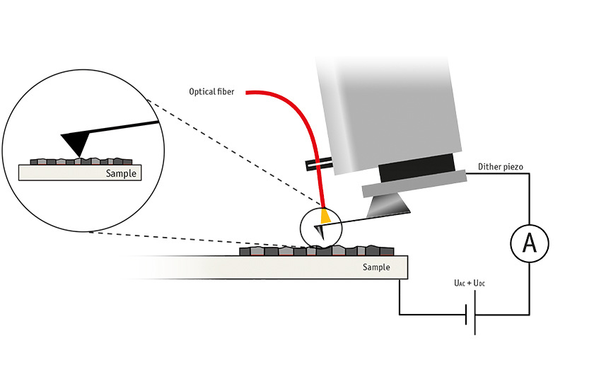
Kelvin probe force microscopy (KPFM) on graphene Graphene belongs to the category of 2D materials and is of interest in the research and development of new devices and materials. Besides atomic resolution imaging of graphene to learn more about crystal orientation, edges and defects, the functional properties of graphene are also of great interest.

Afm Scanning Modes Max Iv
Atomic Force Microscopy (AFM) is a technique that can directly image single molecules in solution and it therefore provides a powerful tool for obtaining unique insights into the basic properties of biological materials and the functional processes in which they are involved.
Contact Mode And Tappingmode Atomic Force Microscopy
The Atomic Force Microscope (AFM) allows for 3D characterization of nanoparticles with sub-nanometer resolution. Nanoparticle characterization using Atomic Force Microscopy has a number of advantages over dynamic light scattering, electron microscopy and optical characterization methods.
Current Distribution Mapping Of Carbon Nanotube Embedded Polymer Using Conductive Atomic Force Microscopy 2020 Wiley Analytical Science
Atomic force microscopy is arguably the most versatile and powerful microscopy technology for studying samples at nanoscale. It is versatile because an atomic force microscope can not only image in three-dimensional topography, but it also provides various types of surface measurements to the needs of scientists and engineers.
Afm Principle How Afm Works Youtube
To learn more about Atomic Force Microscopy, click through. AFM microscopes are among the best solutions for measuring the nanoscale surface metrology and material properties of samples. A conventional compound light microscope is limited to a maximum sample magnification of approximately 1000x; a quantity that is dictated by the wavelengths of ...

Block Diagram Of Atomic Force Microscopy Operation Download Scientific Diagram
Atomic force microscopy should be able to resolve atoms through changes in short-range chemical forces, but resolution is lost if the scanning tip undergoes modifications or if it moves the molecule. Gross et al. (p. 1110) show that in situ functionalization of the tip—for example, with CO—can dramatically improve the resolution of images ...

Atomic Force Microscopy Afm Anton Paar Wiki
Atomic Force Microscope. The atomic force microscope (AFM) is one of the most powerful techniques to investigate the status of surface conditions of either thin film or single crystals, which are being to employ for solar cell applications. ... Schematic diagram showing the principle of atomic force imaging is shown in Fig. 4.1a. As the tip is ...

Atomic Force Microscopy Fig2 Schematic Of Afm Operation
Functionalizing the tip of an atomic force microscope with a CO molecule enabled atomic-resolution imaging of single molecules, and measurement of their adsorption geometry and bond-order relations. In addition, by using scanning tunneling microscopy and Kelvin probe force microscopy, the density of the molecular frontier orbitals and the ...
Bone Biology And Mechanics Lab Bbml
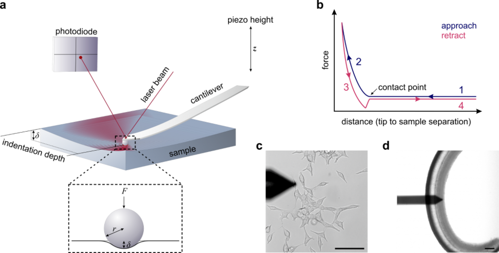
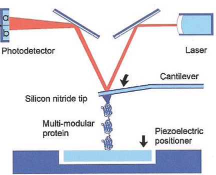
a Single-Molecule Level by Atomic Force Microscopy 179 A B Fig. 2. AFM force spectroscopy: (A) Diagram of a single stretching experiment; (B) Ex ample of a force extension curve, with definitions of force and extension. (Adapted and modified from reference 4, with permission) off the back of the cantilever onto the center of the PSPD.
File Atomic Force Microscope Block Diagram Png Wikimedia Commons
Functionalizing the tip of an atomic force microscope with a CO molecule enabled atomic-resolution imaging of single molecules, and measurement of their adsorption geometry and bond-order relations. In addition, by using scanning tunneling microscopy and Kelvin probe force microscopy, the density of the molecular frontier orbitals and the ...

Afm Witec Raman Imaging
Scanning probe microscopy has been the engine of characterization in nanoscale systems ().Atomic force microscopy (AFM) in particular has developed into a leading technique for high-resolution studies without material restrictions (3-5).It is increasingly being used for detailed characterization in a wide variety of physical, biological, and chemical processes (6, 7).
Atomic Force Microscopy
In this article we will discuss about the design of atomic force microscope, explained with the help of a diagram. An atomic force microscope instead of using a lens is provided with a probe to examine the surface of a specimen with a sharp tip which may be several micrometers in and less than 10 nm in diameter at the point near field to be examined. The tip lies at the end of the lever which ...

Schematic Of An Atomic Force Microscope Download Scientific Diagram

Fundamental Theory Of Atomic Force Microscopy
Sensors Free Full Text Progress In The Correlative Atomic Force Microscopy And Optical Microscopy
Atomic Force Microscopy An Overview From Asylum Research

Atomic Force Microscopy Afm For Polymer Characterization And Analysis Youtube
Conducting Tip Atomic Force Microscopy Fundamentals
Atomic Force Microscopy Microworld Reflections

Laporan Wawasan Pasar Global Atomic Force Microscopy Afm 2020 Tren Peluang Pasar Seluruh Dunia Hingga 2026 Mandennews
Atomic Force Microscopy Nanoscience Instruments

Schematic Representation Of An Atomic Force Microscope Afm With The Download Scientific Diagram
Atomic Force Microscopy
Atomic Force Microscope Afm Microbe Notes

0614 Atomic Force Microscopy Medical Images For Powerpoint Powerpoint Shapes Powerpoint Slide Deck Template Presentation Visual Aids Slide Ppt
Measuring Viscoelasticity Of Soft Biological Samples Using Atomic Force Microscopy Soft Matter Rsc Publishing

Explain Construction And Working Of Atomic Force Microscope Applied Physics 2 Shaalaa Com
Atomic Force Microscopy Afm In The Nanotechnology Lab Dummies

Finzi Lab Research
Afm Atomic Force Microscope University Of Greifswald
Paul Hansma S Website
Schematic Of An Atomic Force Microscope Nist
Atomic Force Microscopy Diagram Vector Image Public Domain Vectors

Atomic Force Microscopy Afm For Topography And Recognition Imaging At Single Molecule Level Springerlink

A Schematic Diagram Of An Atomic Force Microscope Download Scientific Diagram

Afm Cellular And Molecular Biomechanics Laboratory
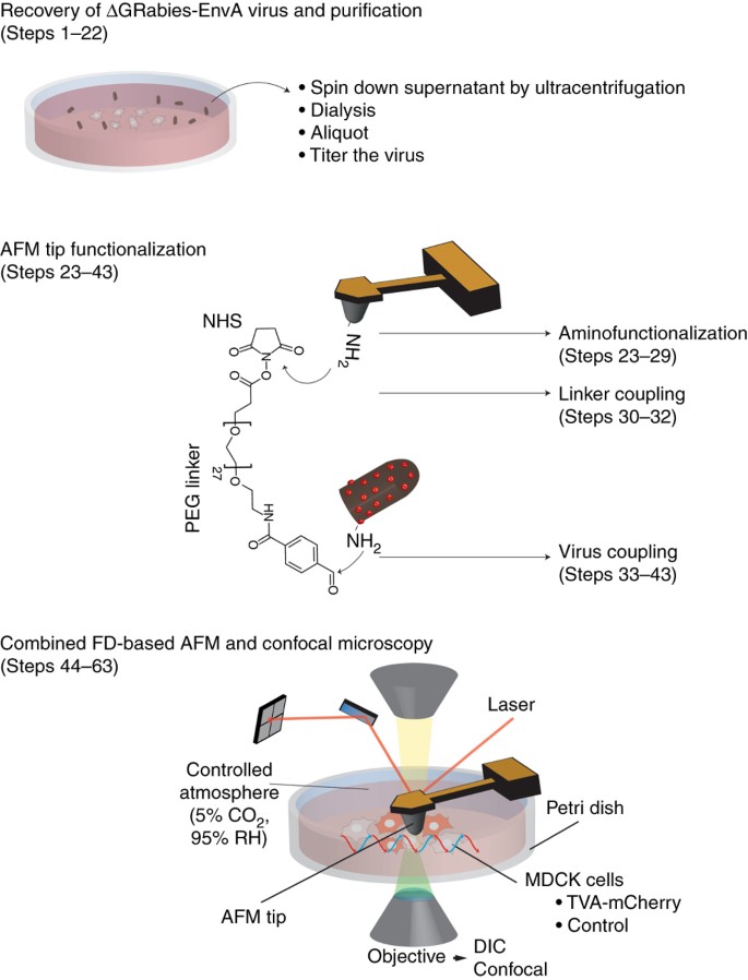
Combining Confocal And Atomic Force Microscopy To Quantify Single Virus Binding To Mammalian Cell Surfaces Nature Protocols
0 Response to "39 atomic force microscopy diagram"
Post a Comment