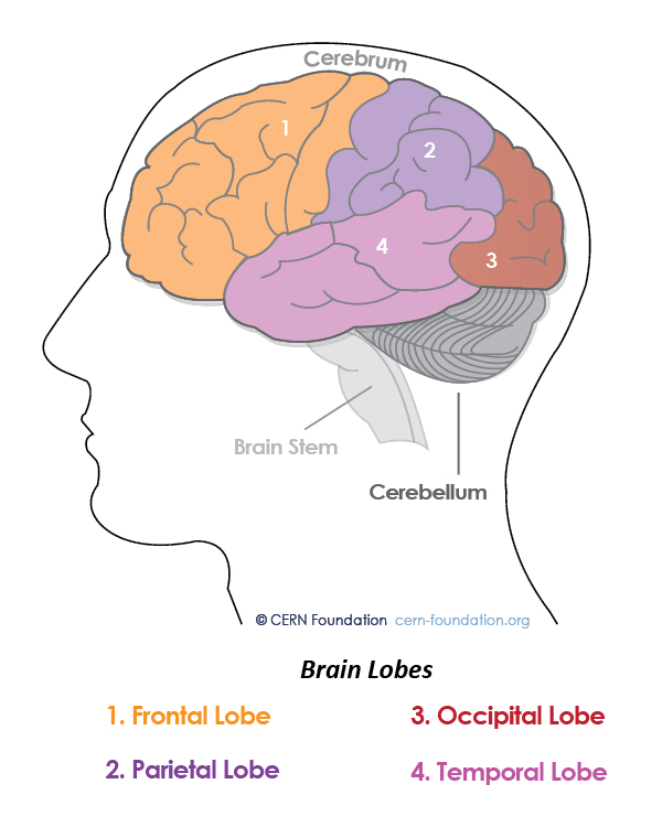
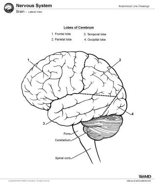
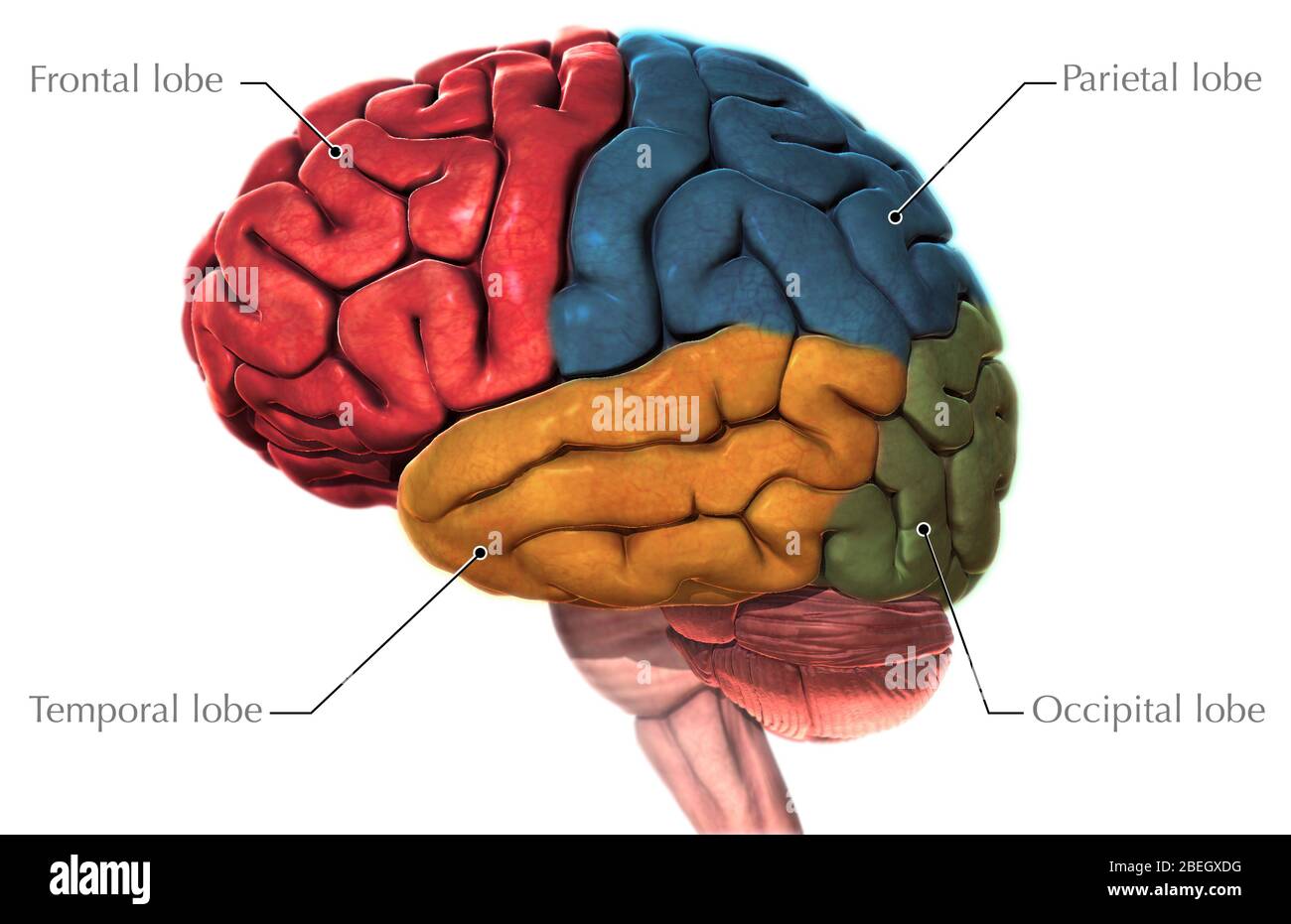
38 lateral view of the brain diagram
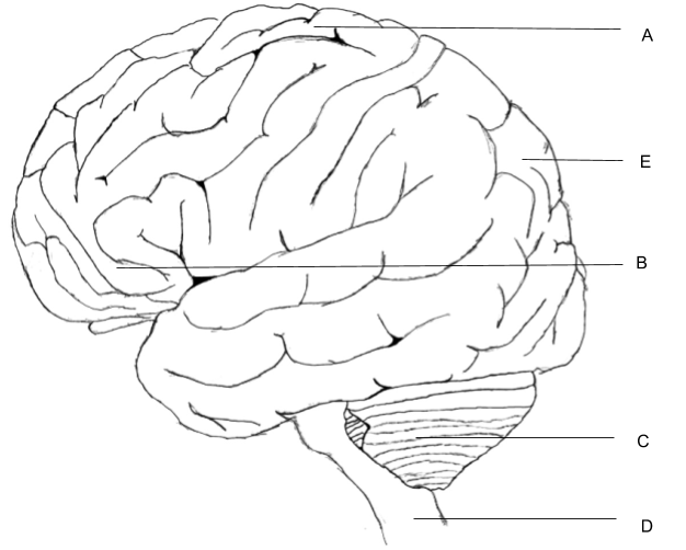
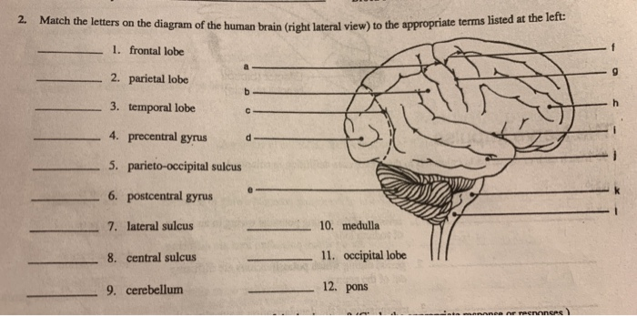
In the diagram of the lateral view of the human brain, parts are indicated by alphabets. Choose the answer in which these alphabets have been correctly matched with the parts which they indicate. If these are the questions swirling in your brain, then this article detailing the diagram of the brain and its functions will definitely whet your appetite regarding brain functions and parts. Of all the human body systems, the nervous system is the most complicated system in the body.
Blank Diagram Complete Diagram. Brain Ventricles: Lateral and Superior Views Blank Diagrams Complete Diagrams Brain Ventricles: 3D Animation The Homunculus . Brainstem/Cranial Nerves Anterior View (Blank Diagram) Anterior View (Complete Diagram) Lateral View (Blank Diagram) Lateral View (Complete Diagram) Cerebral Arterial Circle. Spinal Cord ...
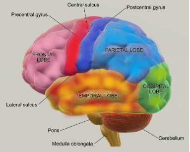
Lateral view of the brain diagram
Label the Brain Anatomy Diagram. The Brain. Read the definitions below, then label the brain anatomy diagram. Cerebellum - the part of the brain below the back of the cerebrum. It regulates balance, posture, movement, and muscle coordination. Corpus Callosum - a large bundle of nerve fibers that connect the left and right cerebral hemispheres. Transcribed image text: XX Figure 7-4 is a diagram of the right lateral view of the human brain (A) Match the letters on the diagram with the following list of terms and insert the appropriate letters in the answer blanks (B) Select different colors for each of the areas of the brain provided with a color-coding circle and use them to color in the coding circles and cor responding structures ... 2,678 labeled brain anatomy stock photos, vectors, and illustrations are available royalty-free. See labeled brain anatomy stock video clips. of 27. brain diagram with labels hypothalamus vector brain diagram pons cerebrum and cerebellum brain pons brain anatomy amygdala brain labelled amygdala brain human midbrain diagram pons.
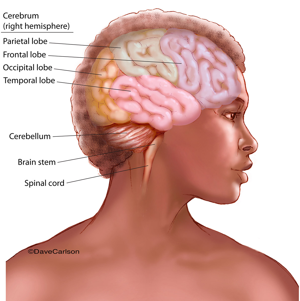
Lateral view of the brain diagram. The forebrain is the anterior part of the brain, which comprises the cerebral hemispheres, the thalamus, and the hypothalamus. It also consists of two subdivisions called the telencephalon and diencephalon. Along with the optic nerves and cranial nerves, the forebrain also includes the olfactory system, or sense of smell as well as the lateral ... Brain. The brain is one of the most complex and magnificent organs in the human body. Our brain gives us awareness of ourselves and of our environment, processing a constant stream of sensory data. It controls our muscle movements, the secretions of our glands, and even our breathing and internal temperature. This is an online quiz called Label Lateral View Of The Brain. There is a printable worksheet available for download here so you can take the quiz with pen and paper. Your Skills & Rank. Total Points. 0. Get started! Today's Rank--0. Today 's Points. One of us! Game Points. 10. Lateral is from the side; medial is towards the midline (often from a sagittal section); dorsal is looking from above; and ventral is looking from below. There are numerous specific parts of the brain that we could name and explore. The following is a list of structures within the four basic subdivisions of the brain: Forebrain
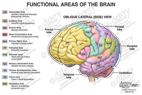
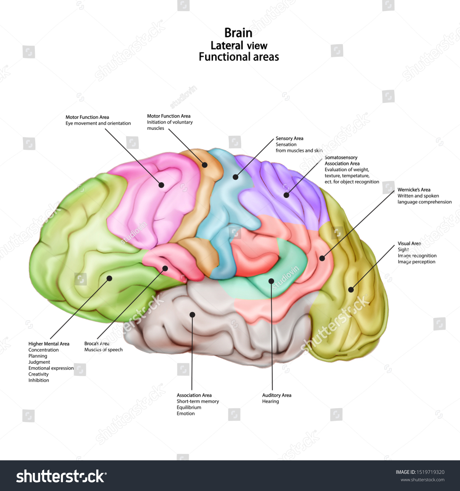
Match the letters on the diagram of the human brain (Gright lateral view) to the appropriate terms listed at the left: lateral view) to 1. frontal lobe 2. parietal lobe 3. temporal lobe 4. precentral gyrusd 5. parieto-occipital sulcus 6. postcentral gyrus 7. lateral sulcus 8. central sulcus 9. cerebellum 10, medulla 11, occipital lobe 12. pons. Download this stock image: Diagram of the lateral view of the human brain, showing the functional areas (motor, sensory, auditory, visual, and speech). - BCE646 from Alamy's library of millions of high resolution stock photos, illustrations and vectors. CENTRAL NERVOUS SYSTEM: Brain. 10. Figure 7-3 is a diagram of the right lateral view of the human brain. Match the letters on the diagram with the following list of terms and insert the appropriate letters in the answer blanks. Color in the corresponding structures in the diagram. If an identified area is part of a . lobe Located lateral to the pyramid of the medulla oblongata; regulates impulse propagation from the cerebrum and midbrain to the cerebellum. Figure 7: Lateral view of the brain stem Marieb & Hoehn (Human Anatomy and Physiology, 9th ed.) - Figure 12.13 Exercise 3:
The frontal lobe is the largest lobe of the brain comprising almost one-third of the hemispheric surface. It lies largely in the anterior cranial fossa of the skull, leaning on the orbital plate of the frontal bone.. The frontal lobe forms the most anterior portion of the cerebral hemisphere and is separated from the parietal lobe posteriorly by the central sulcus, and from the temporal lobe ... 10. This brain is pinned to show the pineal gland, thalamus and lateral ventricle. 11.The image below shows a cleanly separated brain with the major internal structures visible and labeled. 12. Finally, a section of the brain is cut to examine the difference between white matter and gray matter. Want to learn more about the parts of the brain? Try our free brain diagrams and quizzes! Cerebrum. The cerebral cortex, which is the area of the cerebrum seen at a lateral view of the brain, is about 2-5 mm thick and accounts for about 80% of the brain's totalling mass. Its total area has been estimated to be about 2000 cm². Diagram Of The Lateral View Of The Human Brain Showing The Human Brain Brain Art Wernicke S Area. Homunculo Homunculus Brain Homunculus Somatosensory Cortex. Somatosensory Cortex Somatosensory Cortex Hand Wrist Central Nervous System. 14 9 The Cerebrum Contains Motor Sensory And Association Areas Allowing For Higher Mental Functions Primary ...
Complete, labeled illustrations of the parts of the brain in nine different views and sections. It includes detailed diagrams of: brain in place, lateral view, medial view, arteries, frontal section, anterior view, ventricles, horizontal section, and inferior view.
The Brain The Outer Parts of the Brain Divisions of the Brain Actual Human Brain: lateral view, left hemisphere Diagram of Human Brain:lobes and functions Actual Human Brain: Midsection Diagram: Midsection; midbrain and primitive structures Comparison of brains: human; dog; rat Actual Rat Brain Stimulation in the Brains of Animals Split-brain ...
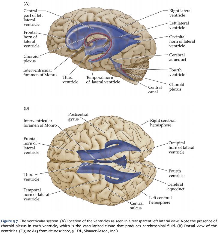
The Brain - Lateral View with Ventricular System. Create healthcare diagrams like this example called The Brain - Lateral View with Ventricular System in minutes with SmartDraw. SmartDraw includes 1000s of professional healthcare and anatomy chart templates that you can modify and make your own.
Figure 12.15b Three views of the brain stem (green) and the diencephalon (purple). View (b) Crus cerebri of cerebral peduncles (midbrain) Infundibulum Pituitary gland Trigeminal nerve (V) Abducens nerve (VI) Facial nerve (VII) Vagus nerve (X) Accessory nerve (XI) Hypoglossal nerve (XII) Pons (b) Left lateral view Glossopharyngeal nerve (IX)
Learn the ventricles of the brain along with their definition, function, location, anatomy, and cerebrospinal fluid (CSF) flow using labeled diagrams. The ventricular system contains the lateral, third, and fourth ventricles whose function is to produce cerebrospinal fluid. Learn where CSF is found,
Brain chart and functions of parts. Brain design and functions of parts. Articles that re so overwhelmed by. Lot of brain diagram and functions of Human Brain Diagram And Functions Anatomy Human Brain, Human Brain and Its Functions, Human Brain Chart, Stress and the Brain Diagram, Diagram of the Human Head Brain Diagram And Functions Brain diagrams can represent the brain in many different ways.
the lateral ventricles) to where it is reabsorbed into the venous blood: Lateral ventricle via openings in the wall of the 4th ventricle surrounding the brain and cord (and central canal of the cord) arachnoid Villi ontaining venous blood 10. Label correctly the structures involved with circulation of cerebrospinal fluid on the accompanying ...
Start studying Lateral View of the Brain. Learn vocabulary, terms, and more with flashcards, games, and other study tools.
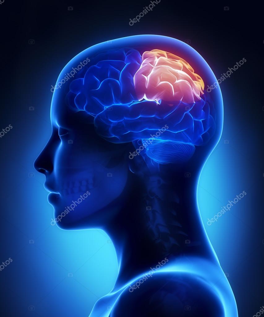
Diagram Of The Lateral View Of The Human Brain, Showing The Functional Areas (Motor, Sensory, Auditory, Visual, And Speech). : News Photo. Save to Board. Save to Board.
Download scientific diagram | A- Lateral view of the human brain and spinal cord. The four sections of the spinal cord are indicated on the left. The nerves that connect the spinal cord to the ...
The Brain - Lateral View - 1. Create healthcare diagrams like this example called The Brain - Lateral View - 1 in minutes with SmartDraw. SmartDraw includes 1000s of professional healthcare and anatomy chart templates that you can modify and make your own.
Ways to View the Brain: the "nervous system" (including the brain) has several orientational directions.(Fisch, 4) It is common to combine terms. For example, a structure may be described as 'dorso-lateral,' which means that it is located 'up and to the side.' (Kolb, 39) A variety of terms is used for different directions and planes of section in the nervous system.
2,678 labeled brain anatomy stock photos, vectors, and illustrations are available royalty-free. See labeled brain anatomy stock video clips. of 27. brain diagram with labels hypothalamus vector brain diagram pons cerebrum and cerebellum brain pons brain anatomy amygdala brain labelled amygdala brain human midbrain diagram pons.

Human Brain Anatomy Set Of Lateral Sagittal Superior Inferior Views With All Lobes Royalty Free Cliparts Vectors And Stock Illustration Image 71810394
Transcribed image text: XX Figure 7-4 is a diagram of the right lateral view of the human brain (A) Match the letters on the diagram with the following list of terms and insert the appropriate letters in the answer blanks (B) Select different colors for each of the areas of the brain provided with a color-coding circle and use them to color in the coding circles and cor responding structures ...
Label the Brain Anatomy Diagram. The Brain. Read the definitions below, then label the brain anatomy diagram. Cerebellum - the part of the brain below the back of the cerebrum. It regulates balance, posture, movement, and muscle coordination. Corpus Callosum - a large bundle of nerve fibers that connect the left and right cerebral hemispheres.

With The Help Of A Labelled Diagram Of Lateral View Of Cerebrum Describe The Structure Biology Shaalaa Com

Cerebrum Lateral Views Organization Of The Brain Cerebrum Superolateral Surface Of Brain Surface Anatomy Of The





/brain_ventricles-56d0ccd03df78cfb37b876dc.jpg)



:background_color(FFFFFF):format(jpeg)/images/library/6721/lateral-views-of-the-brain_english.jpg)


:background_color(FFFFFF):format(jpeg)/images/article/en/lateral-view-of-the-brain/pvf6qv6Bp5acLujnMV4Q_Brain_-_lateral_view.png)
0 Response to "38 lateral view of the brain diagram"
Post a Comment