37 lymph nodes in elbow diagram
28.10.2021 · This article lists a series of labeled imaging anatomy cases by system and modality. Brain CT head: non-contrast axial CT head: non-contrast coronal CT head: non-contrast sagittal CT head: angiogram axial CT head: angiogram coronal CT head... IMAIOS and selected third parties, use cookies or similar technologies, in particular for audience measurement. Cookies allow us to analyze and store information such as the characteristics of your device as well as certain personal data (e.g., IP addresses, navigation, usage …
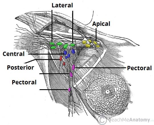
28.10.2021 · Groups of axillary lymph nodes draining the breast (diagram) Axillary lymph nodes. The axillary lymph node chain may be divided into six groups: Apical axillary nodes. Also known as the subclavicular group, they contain 8-12 nodes between the superior border of the pectoralis minor muscle and the clavicle, lateral to the first rib.
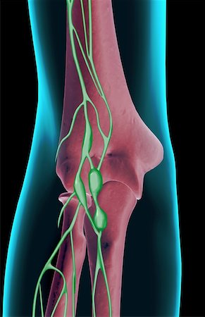
Lymph nodes in elbow diagram
21.10.2021 · Female anatomy diagram: Uterus and ovaries. The uterus is divided into three parts: Body (corpus) - the main part of the uterus, connected to the uterine (fallopian) tubes via the uterine horns.The body has a base (fundus) and an internal chamber (uterine cavity).Isthmus - the constricted part of the uterus, located between the body and the cervix. The lymph nodes of the carcass should be examined if pathological lesions are generalized. Some of the signs of a generalized disease are: Generalized inflammation of lymph nodes including the lymph nodes of the head, viscera and/or the lymph nodes of the carcass. Inflammation of joints. Lesions in different organs including liver, spleen kidneys and heart. The presence of multiple abscesses ... 12.11.2021 · Diagram of axillary lymph node groups. Epitrochlear lymph nodes . Epitrochlear lymphadenopathy is rare, but usually very obvious when present (the patient will often point this out). To assess for epitrochlear lymphadenopathy: 1. Hold the wrist of the side to be examined with your corresponding hand (i.e. right to right). 2. Using your opposite hand, grasp behind the olecranon with …
Lymph nodes in elbow diagram. 13.09.2021 · Finally a diagram summarizes the insertion and origin of the transversal spinalis muscles (semispinalis, multifidus and rotator muscles). NB: the nomenclature of the muscles of the spine varies greatly depending on the source, so we have meticulously followed the Terminologia Anatomica. Anatomy of the cervical vertebral column (Illustration : A. Micheau, MD - E-anatomy - Imaios) Muscles … The breasts are paired structures located on the anterior thoracic wall, in the pectoral region. They are present in both males and females, yet are more prominent in females following puberty. In females, the breasts contain the mammary glands – an accessory gland of the female reproductive system. The mammary glands are the key structures involved in lactation. Top 10 Functions of Ligaments Forming Joints: Joints allow different parts of your body (for example, limbs) to move in different directions. For the formation of a joint, bones need to be joined end to end and fastened with a flexible structure that can allow for movements. 21.04.2021 · The sail sign on an elbow radiograph, also known as the anterior fat pad sign, describes the elevation of the anterior fat pad to create a silhouette similar to a billowing spinnaker sail from a boat. It indicates the presence of an elbow joint effusion.. The anterior fat pad is usually concealed within the coronoid fossa or seen paralleling the anterior humeral line.
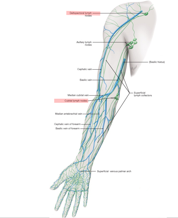
12.11.2021 · Diagram of axillary lymph node groups. Epitrochlear lymph nodes . Epitrochlear lymphadenopathy is rare, but usually very obvious when present (the patient will often point this out). To assess for epitrochlear lymphadenopathy: 1. Hold the wrist of the side to be examined with your corresponding hand (i.e. right to right). 2. Using your opposite hand, grasp behind the olecranon with … The lymph nodes of the carcass should be examined if pathological lesions are generalized. Some of the signs of a generalized disease are: Generalized inflammation of lymph nodes including the lymph nodes of the head, viscera and/or the lymph nodes of the carcass. Inflammation of joints. Lesions in different organs including liver, spleen kidneys and heart. The presence of multiple abscesses ... 21.10.2021 · Female anatomy diagram: Uterus and ovaries. The uterus is divided into three parts: Body (corpus) - the main part of the uterus, connected to the uterine (fallopian) tubes via the uterine horns.The body has a base (fundus) and an internal chamber (uterine cavity).Isthmus - the constricted part of the uterus, located between the body and the cervix.
Elbow Lymph Node Anatomy Stock Photos Page 1 Masterfile
Repository Sustech Edu

Ch 9 Lymphatic System Flashcards Quizlet
1
Axillary Lymph Nodes Definition Anatomy And Location Kenhub

Diagram Of Lymphatic System Showing Lymph Capillaries Lymph Vessels Download Scientific Diagram

Lymph Nodes Anatomy Location Video Lesson Transcript Study Com
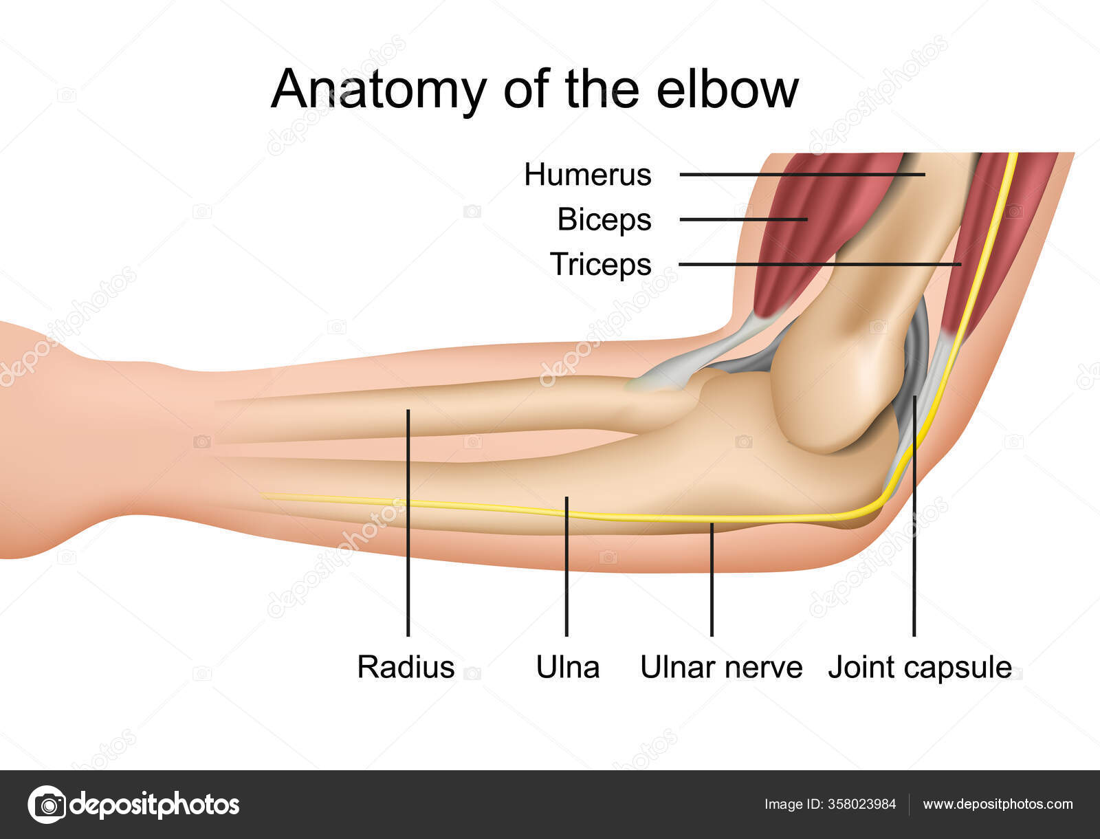
Anatomy Elbow Muscles Medical Vector Illustration Stock Vector Image By C Medicalstocks 358023984

Systematic Anatomy
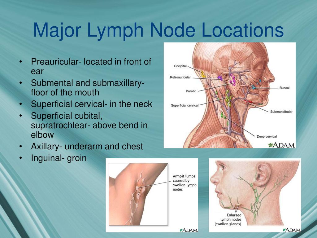
The Lymphatic System The Body S Drains Ppt Download
1
Supratrochlear Lymph Nodes Wikipedia

Lymph Node Exam Protocol Translated To Arabic
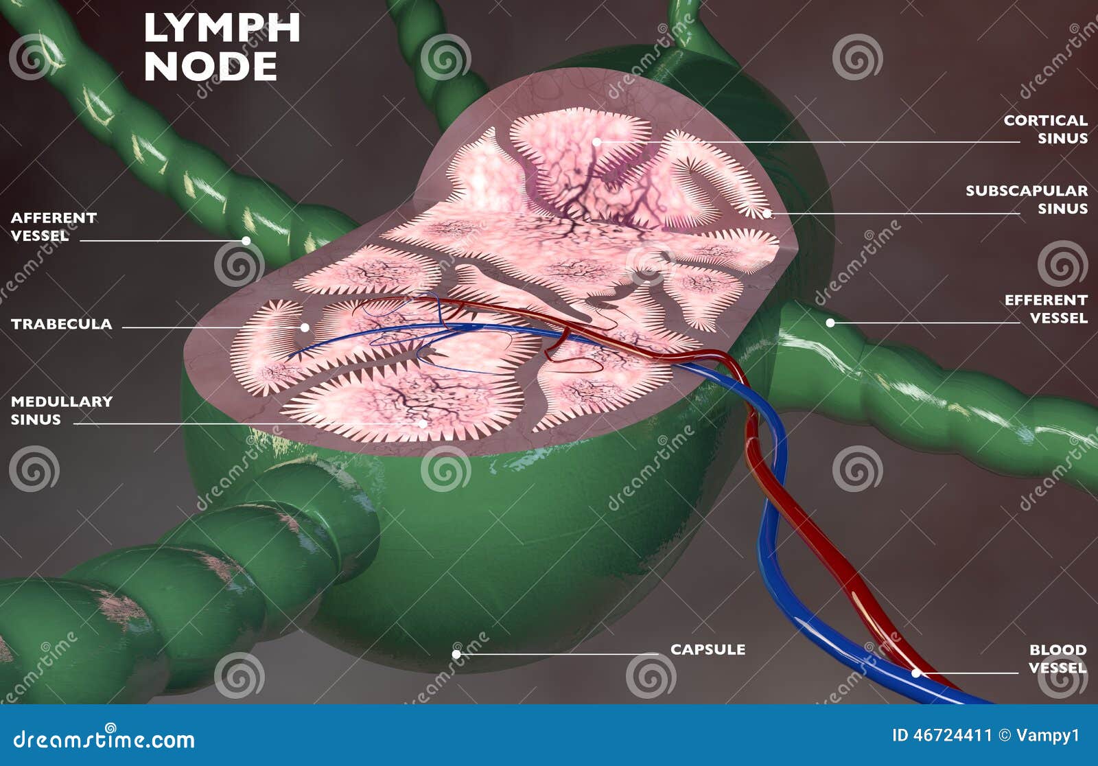
Lymph Node Anatomy Stock Illustrations 1 226 Lymph Node Anatomy Stock Illustrations Vectors Clipart Dreamstime
Elbow Joint Anatomy Video Lecturio Medical
Applied Anatomy Of The Hand Springerlink

Lymph Vessels And Nodes Of Upper Limb Anatomy Intraclavicular Node Lateral Axillary Humeral Nodes Along Axillary Ve In 2021 Lymph Vessels Upper Limb Anatomy Limb

Cubital Lymph Nodes Keyword Search Science Photo Library

Brachial Plexus Png Images Pngwing

3d Rendered Illustration Of A Mans Lymph Nodes Of The Back And Neck Stock Photo Picture And Royalty Free Image Image 117988870

Students Elbow Olecranon Bursitis Information Sinew Therapeutics

Thumb Elbow Vein Anatomy Upper Limb Arm Hand People Anatomy Png Pngwing

Upper Arm And Elbow Knowledge Amboss

Lab Guide 1 Anatomy Of Vessels Bones Of The Extremities 1

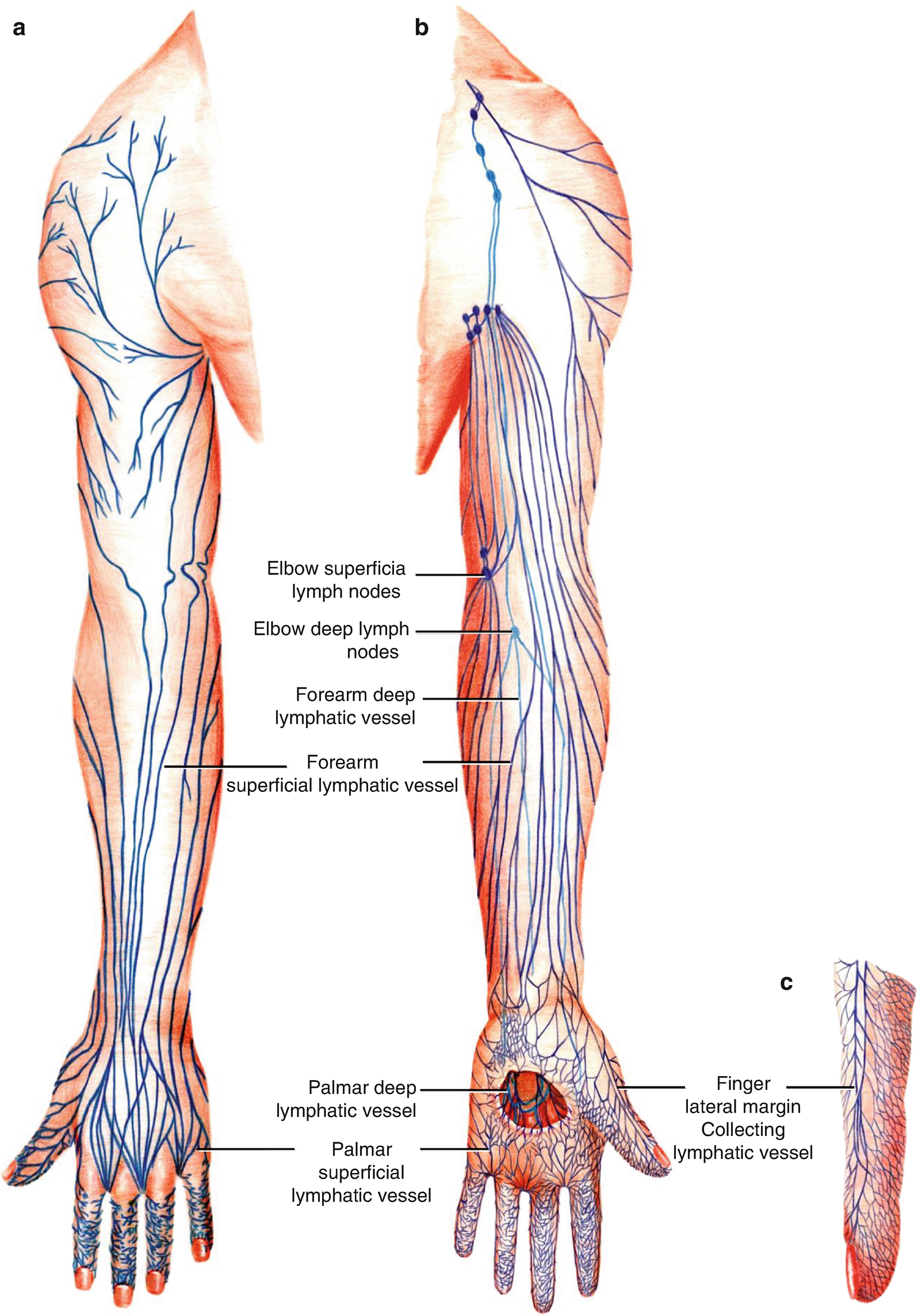
Lymphatic Drainage Of Upper Limb Anatomy
Lymphatic Drainage Of The Upper Limb Vessels Nodes Teachmeanatomy

Vascularized Lymph Node Transfer From The Groin Thoracic Key
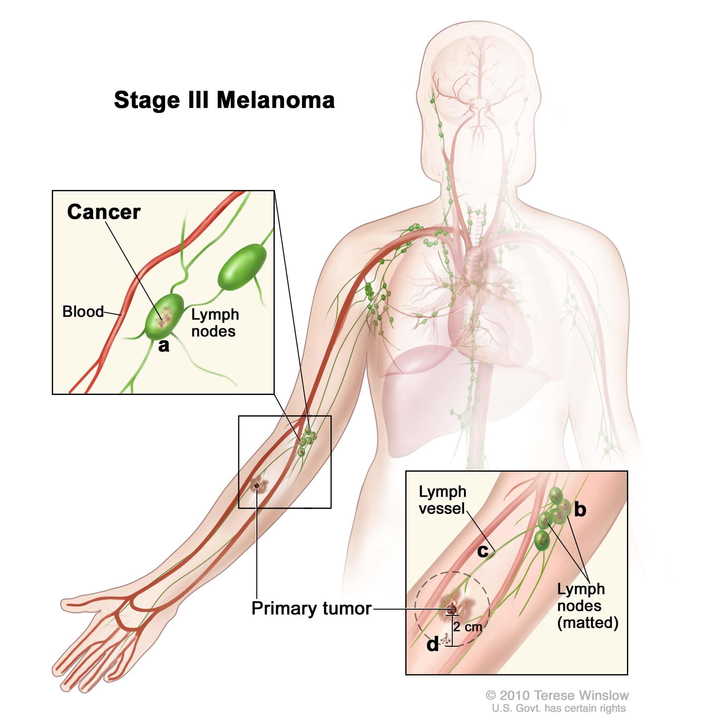
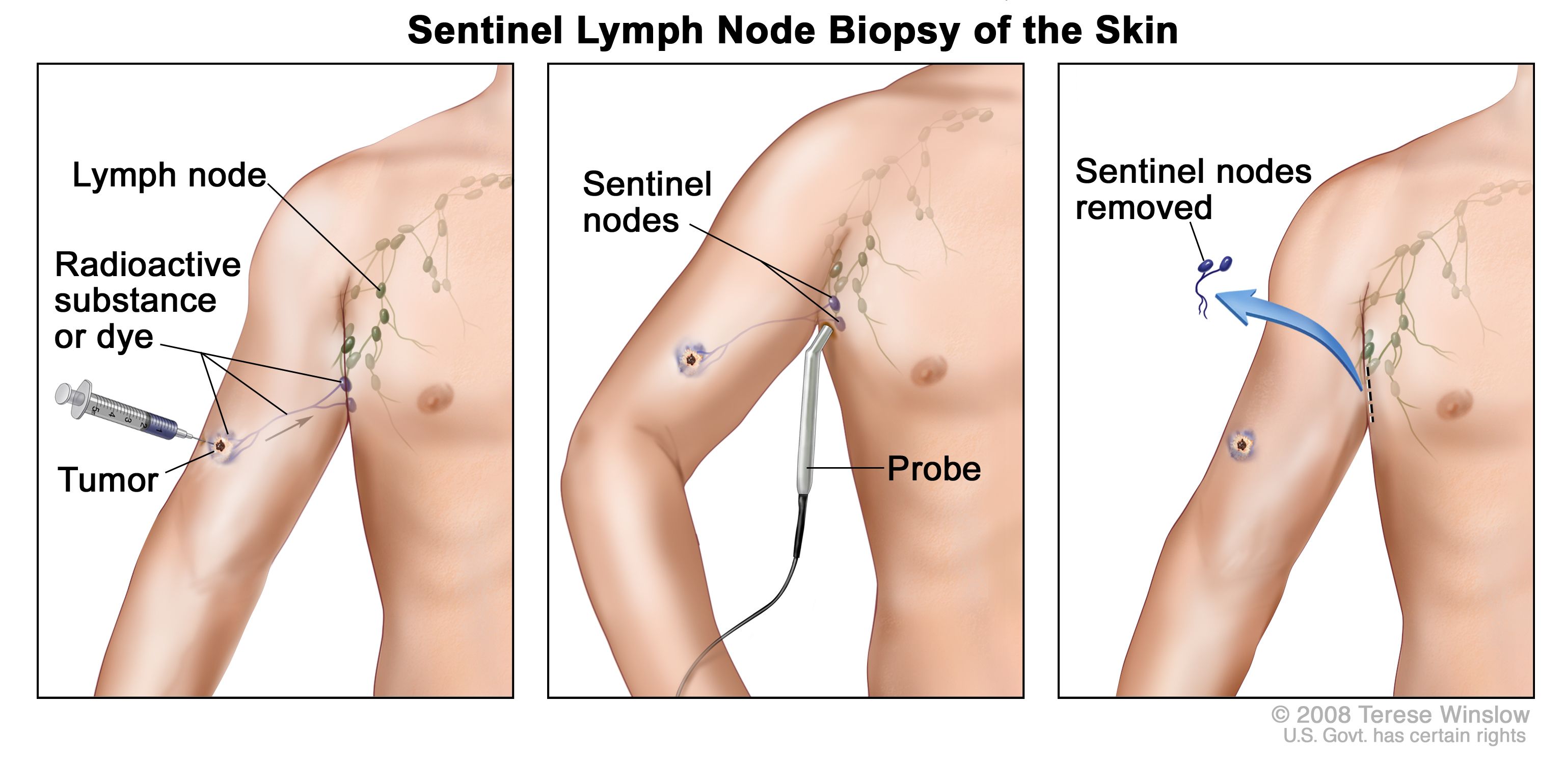
Treatments May Delay Melanoma Recurrence National Cancer Institute

Preoperative View The Scar Of Previous Vascularized Groin Lymph Node Download Scientific Diagram
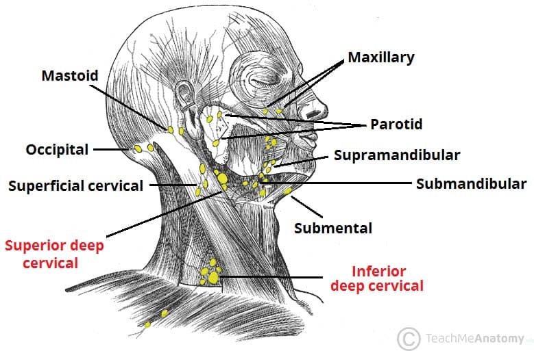
Lymphatic Drainage Of The Head And Neck Teachmeanatomy

Microsurgical Procedures Vascularized Lymph Node Transfer From The Supraclavicular Region Thoracic Key
Superficial And Osseous Upper Limb Flashcards Memorang
1

The Lymphatic System Consists Of The Lymph Conducting Channels Ppt Video Online Download
Merkel Cell Carcinoma Facts Stages Symptoms Dana Farber Cancer Institute Boston Ma

Lymphoreticular Examination Osce Guide Geeky Medics

Us Of The Elbow Indications Technique Normal Anatomy And Pathologic Conditions Radiographics
0 Response to "37 lymph nodes in elbow diagram"
Post a Comment