35 wiggers diagram aortic regurgitation
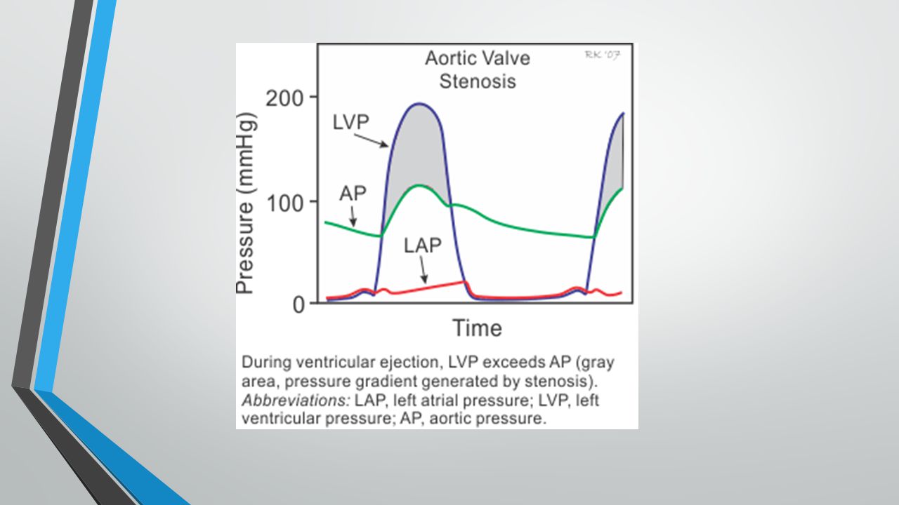
The "Wiggers diagram" ... Mitral and aortic regurgitation are the most common regurgitant lesions. Alternatively, the orifice of a valve may become narrowed or stenotic. This obstructs the flow of blood through it and requires increased pressure gradients to be generated across the valve to achieve Aortic valve stenosis is characterized by the left ventricular pressure being much greater than aortic pressure during left ventricular (LV) ejection (see figure at right). In this example, LV peak systolic pressure during ejection is 200 mmHg (normally ~120 mmHg) and the aortic pressure is slightly reduced to from 120 to 110 mmHg.
This is well-illustrated on a Wiggers diagram where the QRS complex on the ECG directly precedes ventricular systole (represented on the diagram by increased ventricular pressure). Function ... For example, a murmur determined to be systolic rules out diastolic murmur causes such as mitral stenosis and aortic regurgitation. As a general rule ...
Wiggers diagram aortic regurgitation
In early ventricular systole. The semilunar valves open. Ventricular Diastole represents. The period of time the ventricles are filling. Between the end of the T wave and the onset of the QRS complex. Ventricular Diastole. Approximately 70% of the Diastolic filling occurs during. Early Ventricular Diastole. Aortic/Pulmonary artery pressure increases slightly during this phase due to blood pushin back against the now closed aortic/pulmonary valve. Figure 2 : (a) The Wigger's diagram, which shows changes in pressure and volume of heart compartments, as well as electrical and auditory signals that can be detected at different stages of the cardiac ... Early diastolic murmurs immediately follow S2. Examples: aortic and pulmonary regurgitation. Mid-diastolic murmurs (rumble) are due to increased flow (relative stenosis) through the mitral (VSD) or the tricuspid valves (ASD).; Late diastolic murmurs are due to pathological narrowing of the atrioventricular (AV) valves. Example: rheumatic mitral stenosis. Tricuspid stenosis is very rare in childr
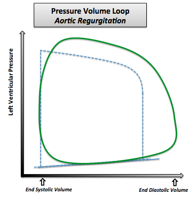
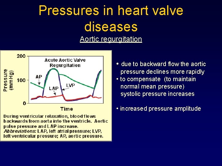
Wiggers diagram aortic regurgitation. Wigger's diagram: This seemingly complex diagram (Don't Panic!) is actually a very useful tool to learn the components of the cardiac cycle and how they relate to cardiac exam findings, which I have indicated in blue. To start, identify the aortic, LV and LA pressure lines and talk yourself through the sequence of Aortic regurgitation Aortic regurgitation Aortic insufficiency (AI) is a condition in which the aortic valve fails to close completely at the end of systolic ejection, causing leakage of blood back through the valve during LV diastole. Cardiac Cycle - Summary and Wigger's Diagram. Aortic valve stenosis Aortic regurgitation occurs when the aortic valve fails to close completely and blood flows back from the aorta (Ao) into the left ventricle after ejection into the aorta is complete and during the time that the left ventricle (LV) is also being filled from the left atrium (LA) (see figure at right). Because the ventricle is being filled from two sources (aorta and LA), this leads to much ... 27. Can we use the term "afterload" for aortic pressure during the phase that the aortic valve is closed? _____ _____ 28. Above diagram shows the relationship of Frank-Starling curves to afterload and stroke volume. The centrally located curve marked "B" is for normal heart. Which of the two curves; A or C, represents increased and ...
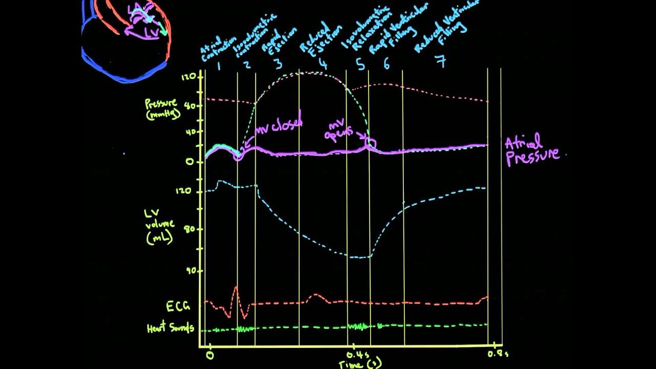
The first set of questions deals with the the events during each of the phases or periods of the normal cardiac cycle. Please use the Wiggers diagrams below showing systemic arterial, left ventricular, and left atrial pressures to answer the questions. In some cases, more than one letter may be circled to achieve the correct answer. FURTHER OBSERVATIONS ON EXPERIMENTAL AORTIC INSUFFICIENCY II. CINEMATOGRAPHIC STUDIES OF CHANGES IN VENTRICULAR SIZE AND IN LEFT VENTRICULAR DISCHARGE By CARLJ. WIGGERS, HAROLD THEISENAND HARLEYA. WILLIAMS (FromtheDepartmentofPhysiology, WesternReserve University MedicalSchool, Clevdand) (Receivedforpublication April30, 1930) INTRODUCTION About Press Copyright Contact us Creators Advertise Developers Terms Privacy Policy & Safety How YouTube works Test new features Press Copyright Contact us Creators ... Wigger Diagram Set-up: Along the bottom, indicate that we'll track time in seconds from 0 to 0.8, which is the duration of a typical healthy cardiac cycle. At the top of the graph, show that we'll track events of the left atrium and left ventricle, as follows: - The left atrium is in systole from time 0 - 0.1, then enters diastole.
position of aortic valve during ventricular filling due to high ventricular pressure. ... changes in Wiggers diagram R heart compared to L heart. all pressures would be lower, valve opening and ventricular volumes would be the same ... regurgitation. murmur often heard throughout systole over the right AV valve. The first diagram shows a side-view of the heart and mitral valve prolapse. Here, in the second diagram, you can see a top-down view of a mitral valve prolapse: Considering the two pictures above…. I guess, according to the cliche, I just saved myself 2,000 words. Phew! Aortic regurgitation. Aortic regurgitation (AR) occurs when there is backflow of blood from the aorta into the left ventricle during ventricular diastole. Aortic regurgitation is associated with an early diastolic murmur heard loudest at the left sternal edge Aetiology. Aortic regurgitation can be either acute or chronic. Chronic AR is often ... The diagram below will show the correct order of assembly. Water Cooled Tig Torch Hook-Up Instructions & Tig Torch Diagram. Before assembling your TIG torch, first consider each of its components. . to set up the water flow properly (See Figure 1 for a diagram). First.A TIG WELDING MACHINE TIGi INTRODUCTION The TIGi is a A inverter-style AC/DC ...
Jcesom Marshall Edu
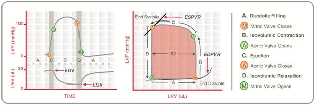
Figure 4.1 Schematic of aortic pressure. Figure 4.2 Wiggers diagram [LVEDV = Left Ventricular End Diastolic Volume; LVESV = Left Ventricular End Systolic Volume]. Figure 4.3 Aortic pressure in a hypovolemic patient. Aortic pressure tracings taken in a patient before and after an intravenous (IV) fluid bolus.
Academic Oup Com
The cardiac cycle is a series of pressure changes that take place within the heart. These pressure changes result in the movement of blood through different chambers of the heart and the body as a whole. These pressure changes originate as conductive electrochemical changes within the myocardium that result in the concentric contraction of cardiac muscle.
5 The Wiggers Diagram Showing Two Cardiac Cycle Events Occurring In Download Scientific Diagram
If a high-pressure jet resulting from aortic stenosis is present, the aorta will stretch If we go back to Part I and look at the Wigger's diagram, the aortic valve is. Dr. Carl Wiggers' careful observations have provided a meaningful resource for students to learn how the heart works. Throughout the many years from his initial .Valvular Stenosis.
1
The Wright table is introduced as a novel tool for teaching and learning the cardiac cycle. It supplements the nearly 100-yr-old Wiggers diagram, which is information rich but difficult for many students to learn. The Wright table offers a compact presentation of information, viewable both in terms of how 1) each compartment's pressures and flows change over time; and 2) the heart works as a ...
Pressure Volume Diagram Wikidoc
midsystolic clicks and the mitral regurgitation heard with mitral valve prolapse o Diastolic Murmurs occur between S2 and S1 Early diastolic murmurs: decrescendo murmurs, arise from aortic or pulmonary valve regurgitation Mid-diastolic murmurs: diamond shaped murmurs, associated with increased

Wigger S Diagram Draw It To Know It
Left Ventricle Pressure Volume Loop. Wiggers' Diagram. Jugular Venous Pressure. Aortic Stenosis. Aortic Regurgitation. Mitral Stenosis. Mitral Regurgitation. Cor Pulmonale. Heart Failure.

Heart Sounds Wikipedia
What elements of the wigger diagram will look different in mitral regurgitation large v wave and rapid y descent A patient is complaining of trouble with breathing - you auscultate and hear a murmur occurring throughout systole, turbulent flow during diastole, and S3.

The Cardiac Cycle Deranged Physiology
Bicuspid valves can trigger heart valve disease including heart valve regurgitation and/or stenosis. Darrell Price says on December 16th, 2008 at 5:55 pm I have an Edwards Bovine aortic valve replacement, procedure done Feb.22, 2007, which is now reguritating.

Progression Of Tricuspid Regurgitation After Surgery For Ischemic Mitral Regurgitation Journal Of The American College Of Cardiology
Read more: Aortic Regurgitation in Chapter 283: Aortic Valve Disease, Harrison's Principles of Internal Medicine 19e. The above Wiggers diagrams are modifications of: adh30 revised work by DanielChangMD who revised original work of DestinyQx; Redrawn as SVG by xavax - Wikimedia Commons: Wiggers Diagram.svg, CC BY-SA 4.0 « What causes alcohol withdrawal seizures?

Valve Defects Cardiac Cycle Ppt Video Online Download
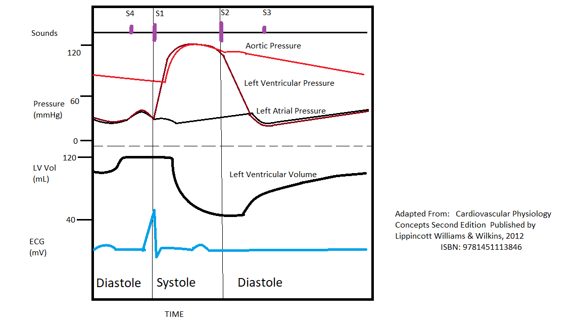
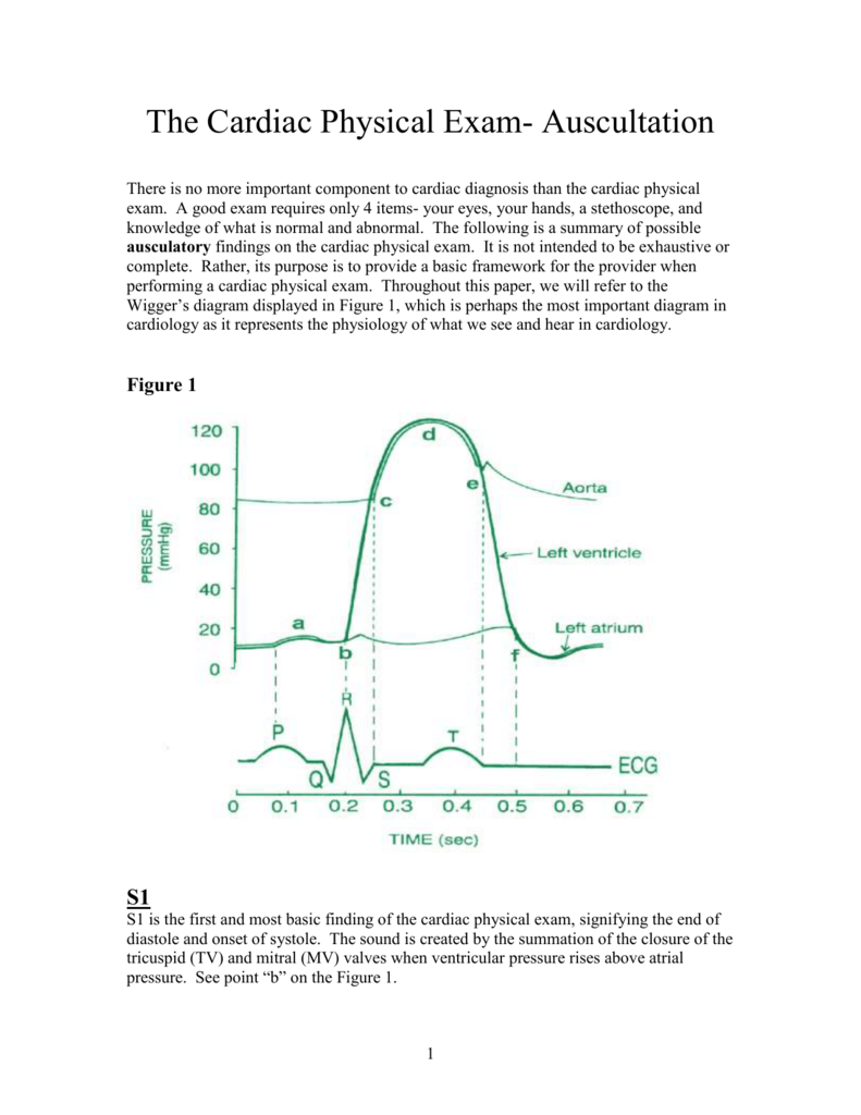
The QRS wave, on the electrocardiogram section of the Wiggers Diagram (image 5.1) represents the beginning of systole. This phase represents ventricular qi depolarization. The rise in pressure in the ventricles exceeds the pressure in the atrium, causing the AV valves to close. The S1 (first heart sound) is the sound heard during this phase. 2.
Pressure Volume Loop Analysis In Cardiology Wikiwand
Wigger's Diagram demonstrating the physiology of diastolic mitral regurgitation. The diagram shows the rapid decrease in diastolic aortic pressure and increase in left ventricular pressure caused by severe aortic regurgitation.

Aortic Regurgitation Knowledge Amboss
Early diastolic murmurs immediately follow S2. Examples: aortic and pulmonary regurgitation. Mid-diastolic murmurs (rumble) are due to increased flow (relative stenosis) through the mitral (VSD) or the tricuspid valves (ASD).; Late diastolic murmurs are due to pathological narrowing of the atrioventricular (AV) valves. Example: rheumatic mitral stenosis. Tricuspid stenosis is very rare in childr
Normal Circulation And Congestive Heart Failure
Aortic/Pulmonary artery pressure increases slightly during this phase due to blood pushin back against the now closed aortic/pulmonary valve. Figure 2 : (a) The Wigger's diagram, which shows changes in pressure and volume of heart compartments, as well as electrical and auditory signals that can be detected at different stages of the cardiac ...

Hemodynamics In The Cardiac Catheterization Laboratory Of The 21st Century Circulation
In early ventricular systole. The semilunar valves open. Ventricular Diastole represents. The period of time the ventricles are filling. Between the end of the T wave and the onset of the QRS complex. Ventricular Diastole. Approximately 70% of the Diastolic filling occurs during. Early Ventricular Diastole.

Pathophysiology Of The Aortic Regurgitation Doppler Signal End Diastolic Notching A Dip Insight Giannakopoulos 2020 Echocardiography Wiley Online Library

Cv Physiology Valvular Insufficiency Regurgitation

Cardiac Cycle An Overview Sciencedirect Topics

Cardiac Physiology For Radiologists Review Of Relevant Physiology For Interpretation Of Cardiac Mr Imaging And Ct Radiographics

Valve Disease Flashcards Chegg Com
Adult Echocardiography Review Ppt Download
Physiology Cardiac Cycle Article

29 Rcis Waveforms Ideas Cardiac Nursing Icu Nursing Pulse Pressure
Mitral Valve Prolapse

Chronic Kidney Disease And Valvular Heart Disease Conclusions From A Kidney Disease Improving Global Outcomes Kdigo Controversies Conference Kidney International

Wiggers Diagram Wikipedia
Examinations In Cardiology I Hemodynamics Jan Ivn Martin

Cardiopulmonology Themedicallyinclined

What Makes The Specific Sound For Each Murmur Crescendo Decrescendo Decrescendo R Step1
Venous Pulse
Cardiac Pe

File Wiggers Diagram Svg Wikimedia Commons
Khan Academy Mcat Vid 2 Pressures In The Cardiac Cycle Youtube Cardiac Cycle Cardiac Nursing Khan Academy

Cv Week 3 Flashcards Quizlet

Coronary Hemodynamics In Patients With Severe Aortic Stenosis And Coronary Artery Disease Undergoing Transcatheter Aortic Valve Replacement Implications For Clinical Indices Of Coronary Stenosis Severity Jacc Cardiovascular Interventions

Wiggers Diagram And Diastolic Pressure R Cardiology
0 Response to "35 wiggers diagram aortic regurgitation"
Post a Comment