37 skin diagram to label
Skin Anatomy: The Layers of Skin and Their Functions The epidermis is made up of five individual layers: 2. Stratum basale: This bottom layer, also known as the basal cell layer, has column-shaped cells that push older cells toward the surface. As the cells move upward, they start to flatten and die. The layer is also made up of melanocytes (that produce a pigment that gives the skin its color ... Skin Diagram || How to draw and label the parts of skin ... 'Skin Diagram || How to draw and label the parts of skin' is demonstrated in this video tutorial step by step.The sense of touch had received supreme importa...
Skin Histology Slide Identification - Thick and Thin Skin ... I hope these skin microscope slide labeled diagrams might help you to identify and learn all the structures. If you need more skin microscope slide labeled diagram, please follow anatomy learner on social media. I will update or upload a new skin slide labeled diagram on social media (if any correction). Functions of skin

Skin diagram to label
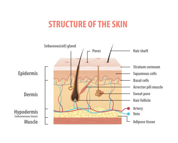
A Human Body Skin-structure Quiz! - ProProfs Sequential Easy First Hard First. Play as. Quiz Flashcard. In this, a human body skin structure quiz, we are going to focus on the underlying and the most elementary structure of the human body. It's easy to take your skin for granted, but when you consider how it protects your body from harm, it is something we should appreciate more. The Skin (Human Anatomy): Picture, Definition, Function ... The skin is the largest organ of the body, with a total area of about 20 square feet. The skin protects us from microbes and the elements, helps regulate body temperature, and permits the ... byjus.com › biology › skin-diagramSkin Diagram with Detailed Illustrations and Clear Labels Skin Diagram. The largest organ in the human body is the skin, covering a total area of about 1.8 square meters. The skin is tasked with protecting our body from the external elements as well as microbes. The skin is also responsible for maintaining our body temperature – this was apparent in victims who were subjected to the medival torture ...
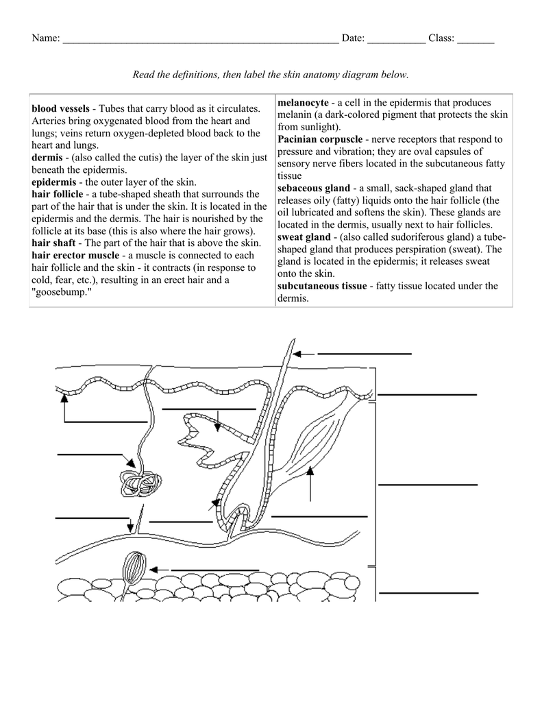
Skin diagram to label. Label_the_Skin_Anatomy_Diagram_AP_online.docx - Name_ Date ... Name_____ Date _____ Period_____ Label the Skin Anatomy Diagram Read the definitions, then label the skin anatomy diagram below. blood vessels - Tubes that carry blood as it circulates. Arteries bring oxygenated blood from the heart and lungs; veins return oxygen-depleted blood back to the heart and lungs. dermis - (also called the cutis) the layer of the skin just beneath the epidermis ... Label The Skin Anatomy Diagram Answers Label The Skin Anatomy Diagram Answers 1/4 [EPUB] Label The Skin Anatomy Diagram Answers Anatomy and Physiology-J. Gordon Betts 2013-04-25 Hole's Essentials of Human Anatomy and Physiology-David Shier 2006-01-01 Designed for the one-semester anatomy and physiology course, "Hole's Essentials of Human Anatomy and Physiology" assumes no prior science knowledge and supports core topics with ... Skin Diagram Worksheets & Teaching Resources | Teachers ... 1. $4.00. PDF. Product DescriptionThis resource includes all the information you will need to help your students label the basic structures of the skin. It also includes two versions of the oversized diagram, as well as basic notes and a Exploration Guide to accompany the diagram. My students struggle because most. PDF Anatomy of Skin - Lancaster High School referred to as thin skin. In areas of the body exposed to greater friction, like the fingertips, palms and soles of the feet the epidermis ... Look at the diagrams on pg 103 in the red textbook to help create your posters. Summary On a loose-leaf sheet of paper with you name on it, answer the following: ...
› skin › labelLabel Skin Diagram Printout - EnchantedLearning.com Read the definitions, then label the skin anatomy diagram below. blood vessels - Tubes that carry blood as it circulates. Arteries bring oxygenated blood from the heart and lungs; veins return oxygen-depleted blood back to the heart and lungs. dermis - (also called the cutis) the layer of the skin just beneath the epidermis. Integumentary system parts: Quizzes and diagrams | Kenhub Labeled diagram of the skin. So what's the idea? Spend some time analyzing the skin diagram labeled above. Try to memorize the appearance and location of each structure. Learning the function of each structure will accelerate your ability to memorize, so be sure to check out our detailed article on The Integumentary System parts and functions. Skin Labeling | Biology Game | Turtle Diary Identify and label figures in Turtle Diary's interactive online game, Skin Labeling! Drag the given words to the correct blanks to complete the labeling! healthiack.com › encyclopedia › skin-diagram-labeledSkin diagram labeled - Healthiack Best viewed on 1280 x 768 px resolution in any modern browser. Skin diagram labeled 1075. Skin diagram labeled 1077. Skin diagram labeled 1080. Skin diagram labeled 1082. Skin diagram labeled 1087. Skin diagram labeled 1089. Skin diagram labeled 1097. Skin diagram labeled 1111.
quizlet.com › 534698122 › labeled-skin-structure-diagramLabeled Skin Structure Diagram | Quizlet Labeled Skin Structure. STUDY. Learn. Flashcards. Write. Spell. Test. PLAY. Match. Gravity. Created by. Styson64 TEACHER. Figure 4.3 from pg 114 of your textbook. Terms in this set (20) ... Fibrous and elastic tissue, provides strength and elasticity to the skin and supports the epidermis, home to hair follicles, glands, nerves etc. PDF Title: Skin Structure - Kent State University Beginning students may work with a partner or use their notes to help them label the diagram. Adaptations for Advanced Students Advanced students may research some additional parts of the skin (such as melanocyte, melanin, or corpuscles) or a related topic (such as sunburn, skin cancer, or moles and freckles) to explain to the class. Label the Skin Quiz - PurposeGames.com This is an online quiz called Label the Skin. There is a printable worksheet available for download here so you can take the quiz with pen and paper. Your Skills & Rank. Total Points. 0. Get started! Today's Rank--0. Today 's Points. One of us! Game Points. 11. You need to get 100% to score the 11 points available. Structure and Functions of Skin - Anatomy, Diagram and ... The skin has a surface area of between 16.1-21.5 sq ft. for an adult human. The thickness of the skin differs over all parts of the body, and between men and women and the young and the old. For example, the skin on the forearm which is on average 1.3 mm in the human male and 1.26 mm in the human female.
Medical Terminology 4f: Integumentary System - Label the ...
Label The Skin Anatomy Diagram Tag Human Skin Diagram ... This article will look at the components and the accessory structures of the integumentary system, skin healing, skin integrity, and the staging of pressures ulcers. This article contains 7 Facts about the Integumentary System Every Nursing Student Should Know. #nursecepts #iintegumentarysystem #nursingstudent #nursingschool

Assignment 11 pg 104.pdf - 4. Label the skin structures and ...
› cms › lib04Skin Diagram Labeling - New Providence School District Skin Diagram Labeling . 1. Label the diagram with the . letters. below according to the structure/area they describe. You may label with a line or put the label directly onto the area described. Be as precise as possible. If you are worried about the precision of your label add a word after to explain exactly where your label should be.

Skin Appendages & Skin Cancer - ppt download
Label Skin Diagram Worksheet - Diy Color Burst Skin Diagram Labeling. Add labels to the diagram of the skin shown below. Worksheet December 18 2017. Label skin diagram worksheet. Which of the following happens to epidermal cells as they move up to the surface of the skin. The dermis the epidermis fat layer. The outermost layer of the skin is.
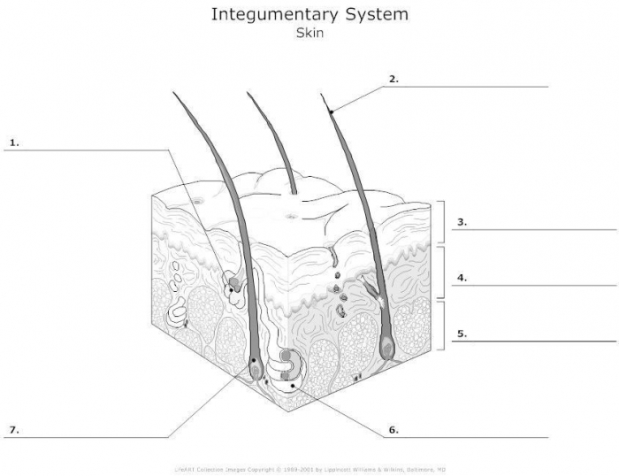
Skin Diagram Labeling
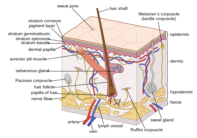
Anatomy of the Skin | SEER Training The skin is the body's largest organ; covering the entire outside of the body, it is about 2 mm thick and weighs approximately six pounds. It shields the body against heat, light, injury, and infection. The skin also helps regulate body temperature, gathers sensory information from the environment, stores water, fat, and vitamin D, and plays a ...
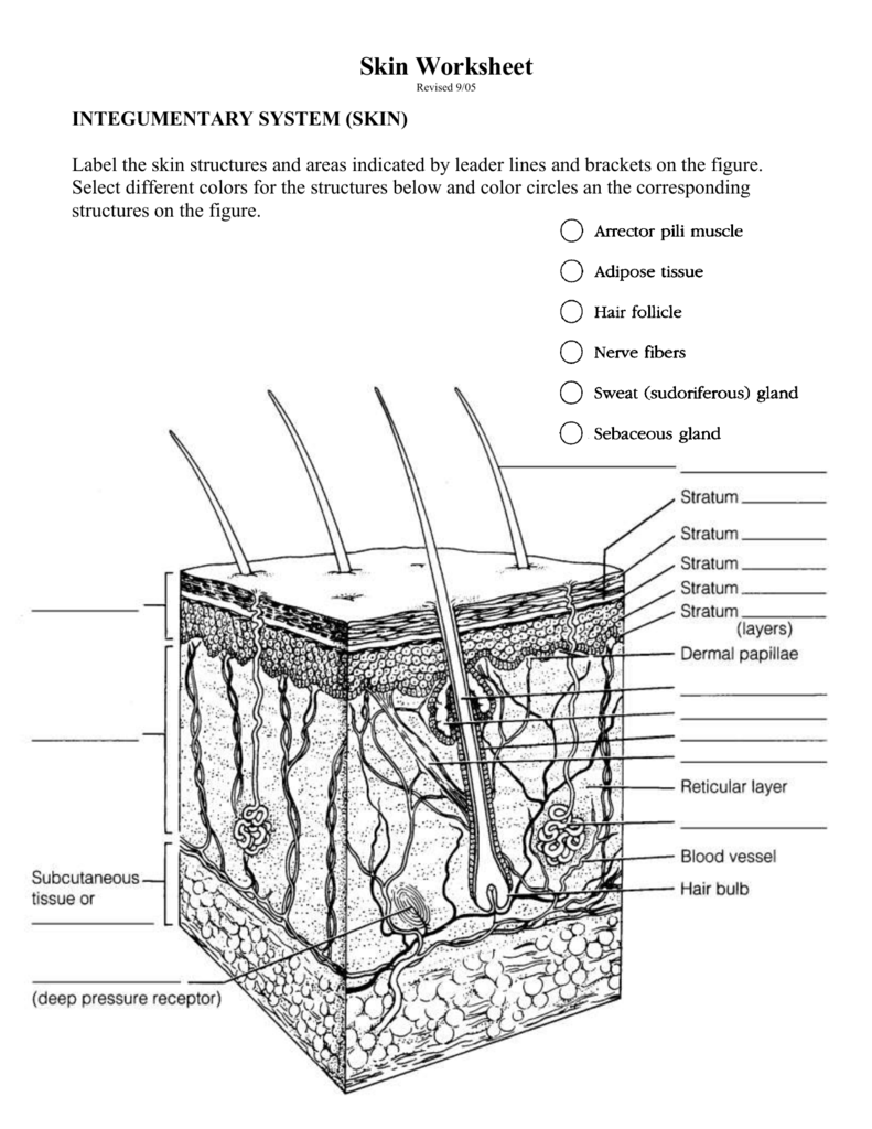
Skin Worksheet
Skin Diagram and Information About Your Skin This is the deepest of the layers of skin, and is located on the bottom of the skin diagram. It connects or binds the dermis above it to the underlying organs. This layer is mainly composed of loose fibrous connective tissue and fat (adipose) cells interlaced with blood vessels. In females, the hypodermis is generally about 8% thicker than in ...

Label the Skin Quiz
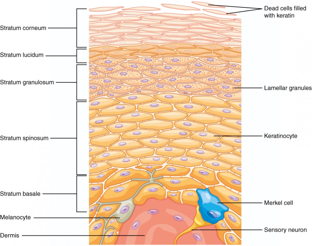
PDF Integumentary System Part I: Functions & Epidermis - found only in thick skin - (this is what your book says. Not true, this layer is everywhere, it is just thinner in other parts, unless abrasion occurs then it thickens.) Stratum Corneum • The "horn layer": - exposed surface of skin - shed and replaced every 2 weeks - cells constantly flake off • feeds the dust mites in your ...
Human skin - Wikipedia
Skin diagram labeled | Healthiack Skin diagram labeled 1077. This image displays Skin diagram labeled.

Skin 2: accessory structures of the skin and their functions ...
Skin Model (labeled) Diagram | Quizlet Start studying Skin Model (labeled). Learn vocabulary, terms, and more with flashcards, games, and other study tools.

Help me name the Skin Diagram please - Brainly.com
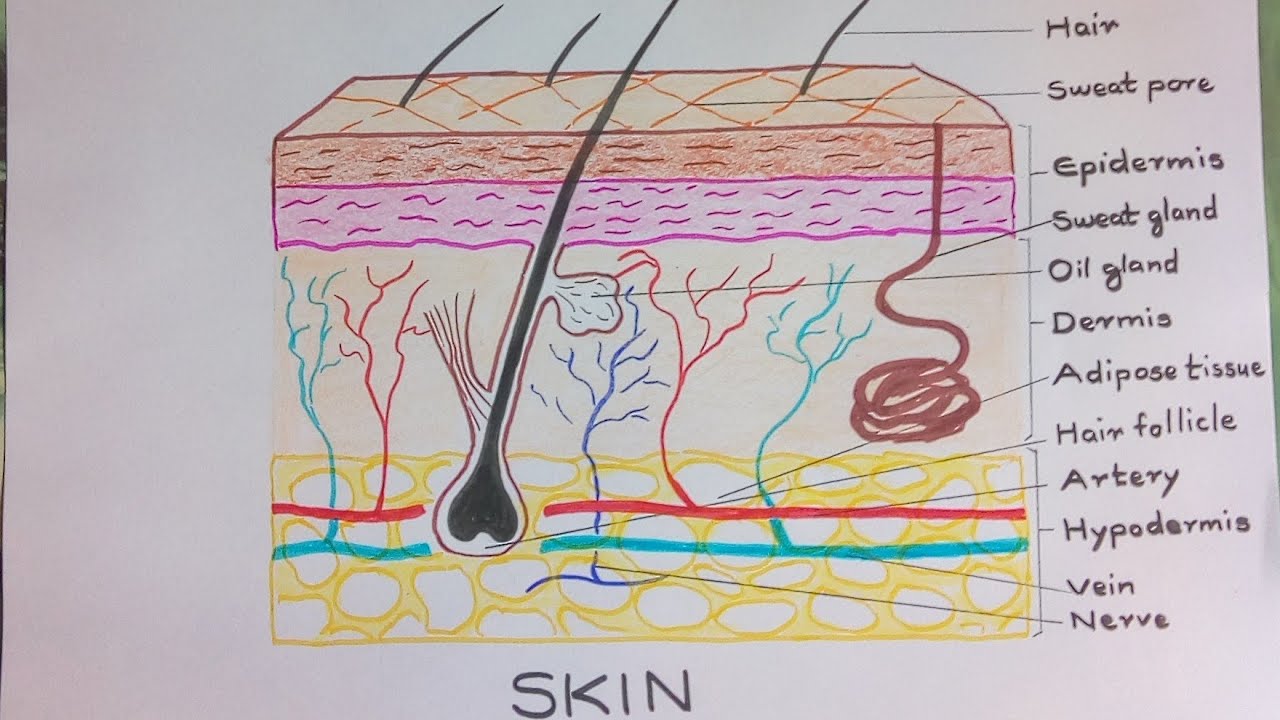
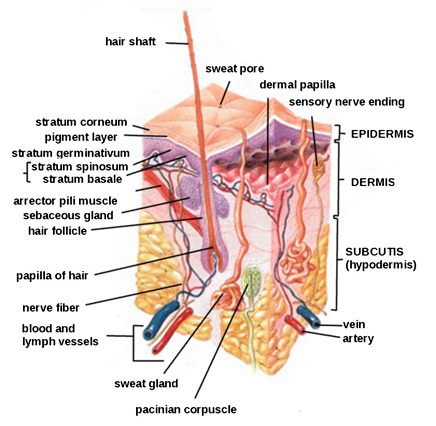
wordwall.net › resource › 5230954Skin diagram to label - Labelled diagram - Wordwall Epidermis, Dermis, Hypodermis, Blood and lymph, Sensory nerve ending, Sweat gland, Arrector pili muscle, Sebaceous gland, Hair shaft, Dermal papilla.

DRAW IT NEAT : How to draw skin LS
Solved Anatomy & Physiology - Integumentary System ... Anatomy and Physiology questions and answers. Anatomy & Physiology - Integumentary System Worksheet - Basic skin structure Name: 1. Label the diagram by placing the correct letter in the box a. hypodermis b. stratum basale c. dermis d. stratum corneum e stratum spinosum f. stratum lucidum g. stratum granulosum h. epidermis i. reticular j ...
Skin-and-Membranes-Worksheet
Label diagram of the skin Quiz - purposegames.com This is an online quiz called Label diagram of the skin. There is a printable worksheet available for download here so you can take the quiz with pen and paper. Your Skills & Rank. Total Points. 0. Get started! Today's Rank--0. Today 's Points. One of us! Game Points. 17. You need to get 100% to score the 17 points available.

Label the Skin Diagram | Quizlet
byjus.com › biology › skin-diagramSkin Diagram with Detailed Illustrations and Clear Labels Skin Diagram. The largest organ in the human body is the skin, covering a total area of about 1.8 square meters. The skin is tasked with protecting our body from the external elements as well as microbes. The skin is also responsible for maintaining our body temperature – this was apparent in victims who were subjected to the medival torture ...

Labeling the Parts of Skin Diagram | Quizlet
The Skin (Human Anatomy): Picture, Definition, Function ... The skin is the largest organ of the body, with a total area of about 20 square feet. The skin protects us from microbes and the elements, helps regulate body temperature, and permits the ...
Label the skin - Teaching resources
A Human Body Skin-structure Quiz! - ProProfs Sequential Easy First Hard First. Play as. Quiz Flashcard. In this, a human body skin structure quiz, we are going to focus on the underlying and the most elementary structure of the human body. It's easy to take your skin for granted, but when you consider how it protects your body from harm, it is something we should appreciate more.

Integumentary System Labeling Diagram | Quizlet
19,704 Skin Anatomy Stock Photos, Pictures & Royalty-Free ...

Skin Diagram | Worksheet | Education.com
Skin Diagram || How to draw and label the parts of skin
Label a diagram of the skin - Mrs. Sanborn's Science Class

Given below is a diagrammatic sketch of the vertical section ...
Answers: Label the Skin Diagram 피부 해부도

Accessory Structures of the Skin | Anatomy and Physiology

Human Body: The Skin

Anatomy of Skin art print poster
(118).jpg)
A Human Body Skin-structure Quiz! - ProProfs Quiz
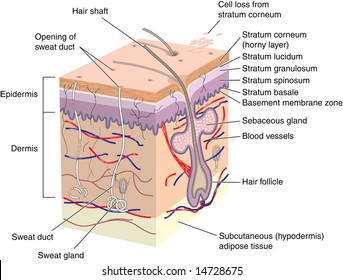
Cross Section Human Skin Labels Stock Illustration 14728675

Label The Skin Anatomy Diagram Tag Human Skin Diagram Label ...

A diagrammatic representation of the structure of human skin ...

Skin 1: the structure and functions of the skin | Nursing Times
Integumentary system review key
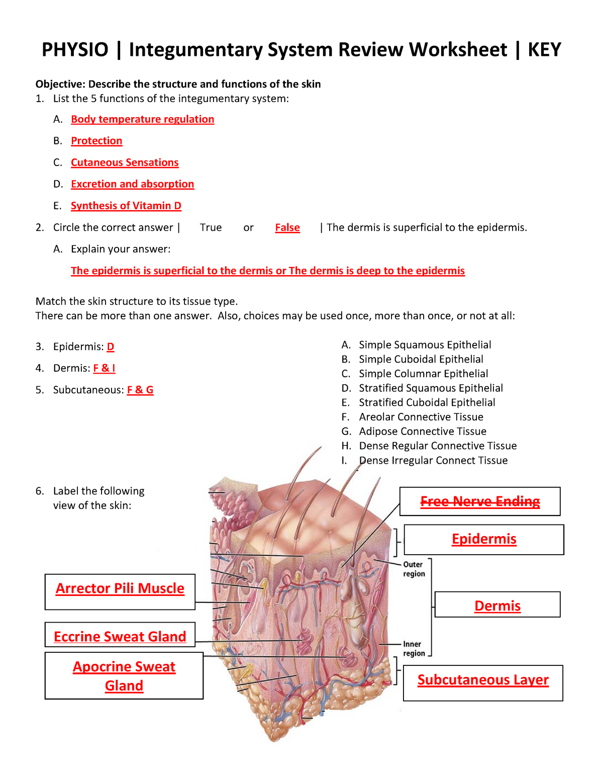
Integumentary System Review Worksheet KEY ( Physio) 2013-2014 ...
Skin Diagram Worksheets | 99Worksheets
Layers of the Skin | Anatomy and Physiology I

Human skin - Wikipedia

SKIN APPENDAGES HAIR NAILS GLANDS September 23 24

Subcutaneous tissue - Wikipedia
Skin Anatomy - EnchantedLearning.com
0 Response to "37 skin diagram to label"
Post a Comment