37 where on the diagram is the femoral area?
Sep 07, 2021 · The dog’s hip bone consists of four distinct bones – ilium, ischium, pubis, and acetabular bone. The left and right ilium bones of a dog are almost parallel to each other. You will find a concave area in the gluteal surface of dog ilium bones. You will also find some other special anatomical features in the hip bones of a dog. Rectum: At the end of the large intestine, this small space is a temporary storage area for feces. Anus: This is the external opening of the rectum, ... Femoral artery. Medically reviewed by Avi Varma, MD. The femoral artery is one of the major arteries in the human body. Its primary function is to supply blood to the lower section of the body.
Being overweight or obese can increase the pressure on your lateral femoral cutaneous nerve. Pregnancy. A growing belly puts added pressure on your groin, through which the lateral femoral cutaneous nerve passes. Diabetes. Diabetes-related nerve injury can lead to meralgia paresthetica. Age. People between the ages of 30 and 60 are at a higher ...

Where on the diagram is the femoral area?
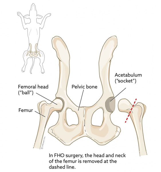
The femoral head forms the "ball" in the ball and socket joint of the hip. It is also located within the joint capsule and is covered by a synovial membrane. Head of femur Caput femoris 1/3 The femoral neck is about 5 cm long and can be subdivided into three regions. 32) Where on the diagram is the femoral area? a) D b) E c) F d) J e) K Answer: a Difficulty: Medium Learning Objective 1: LO 1.5 Describe the anatomical position and how anatomical terms are used to describe the human body. Start studying Arteries & Veins. Learn vocabulary, terms, and more with flashcards, games, and other study tools.
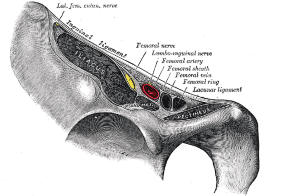
Where on the diagram is the femoral area?. A diagram shows the various inguinal lymph nodes (lymphatic ganglia). The chapter on the innervation of the lower limb presents diagrams of the lumbosacral plexus and its main nerve branches for the lower limb (lateral cutaneous nerve of the thigh, femoral nerve, sciatic nerve and posterior cutaneous nerve of the thigh and obturator nerve). The femoral nerve is the major nerve in your thigh. It's one of the largest leg nerves and runs from your pelvis down the front of your leg. The nerve signals carried by the femoral nerve are a critical part of the ability to stand, walk, and maintain balance. 1 Anatomy Nerves are complex structures that branch out like a tree. Unit 1 Picture questions. Where on the diagram is the femoral area? B) They act as the receptors. Study the diagram. What is the role of the stretch-sensitive nerve cells in the cervix? A) They act as the stimulus. B) They act as the receptors. The femoral artery is one of the major arteries in the human body. It extends from the iliac artery near the abdomen down to the legs. The primary function of this artery is to supply blood to the...
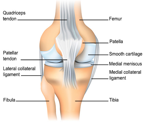
The most common injury to the lateral femoral condyle is an osteochondral fracture combined with a patellar dislocation. The osteochondral fracture occurs on the weight-bearing portion of the lateral condyle. Typically, the condyle will fracture (and the patella may dislocate) as a result of severe impaction from activities such as downhill skiing and parachuting. ... 10.01.2022 · venn diagram of type 1 and type 2 diabetes Ginseng. Used as a traditional medicine for millennia, studies suggest that both Asian and American ginseng may help lower blood sugar in ...Exercise can reduce blood pressure, improve glucose tolerance, and reduce too-high blood sugar levels. The ADA makes the same recommendations for those ... 15.08.2020 · The knee joint is a hinge type synovial joint, which mainly allows for flexion and extension (and a small degree of medial and lateral rotation). It is formed by articulations between the patella, femur and tibia. In this article, we shall examine the anatomy of the knee joint – its articulating surfaces, ligaments and neurovascular supply. It crosses the back of the knee obliquely and inserts on the posterior surface of the upper tibia bone in the lower leg. This muscle is involved in rotation of the knee joint during movements in which the foot is on the ground. The action of the popliteus depends on whether the femur or tibia is in a fixed position.
Thank you for being Super. Just like on a map a region refers to a certain area. Pin On Teaching Ideas Worksheets The nose is referred to as the nasal region. Body regions blank diagram. The left lumbar region consists of the descending colon the. Both the liver and the stomach are located in the […] 18.03.2015 · Mammary glands and ducts are also important in gauging the stage of a woman’s breast cancer. Staging depends on where the cancer is located and what areas are affected. The femoral nerve can be susceptible to damage from pelvic fractures because if the anterior pelvic bones crack, they can press on and possibly sever the femoral nerve. The best way to spot femoral nerve damage is if leg movement becomes impaired (particularly straightening the leg) and there is a lasting numbness in the area. The lumbar plexus is a web of nerves (a nervous plexus) in the lumbar region of the body which forms part of the larger lumbosacral plexus.It is formed by the divisions of the first four lumbar nerves (L1-L4) and from contributions of the subcostal nerve (T12), which is the last thoracic nerve.Additionally, the ventral rami of the fourth lumbar nerve pass communicating branches, …
Hip Picture Image on MedicineNet.com
Diagrams! They remind me of school textbooks which used to have plenty of them, providing a visual aid to understanding difficult subjects. This article explains the nervous system function and structure with the help of a human nervous system diagram and gives you that erstwhile 'textbook feel'.
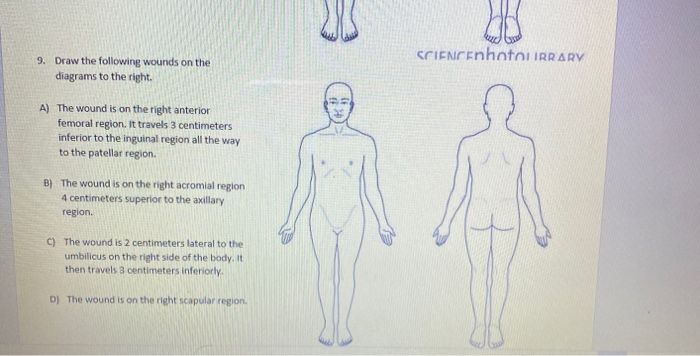
Solved 8. Describe the wounds shown using at least 3 | Chegg.com
In the diagram,the femoral area is _____to the cervical area? A)superior B)inferior C)medial D)proximal E)posterior. Correct Answer: Explore answers and other related questions . 10+ million students use Quizplus to study and prepare for their homework, quizzes and exams through 20m+ questions in 300k quizzes.
What is the anatomy of femoral triangle relevant to femoral ...
The lateral femoral cutaneous nerve is a branch of the lumbar plexus, exiting the spinal cord between the L2 and L3 vertebrae. It emerges at the lateral edge of the psoas muscle group, below the ilioinguinal nerve, and then passes beneath the iliac fascia and the inguinal ligament.

Anatomy of the Femoral Region - TrialExhibits Inc.
In the diagram the femoral area is to the cervical area a superior b inferior c from BIO 2301 at St. John's University
Femoral Region and Hernias: Anatomy - Lecturio Medical
Download scientific diagram | Synthetic femoral models were cut from the middle diaphyseal region with a final size of 200.0 mm. The distal end was firmly fixed with polymethylmethacrylate to each ...

Femoral Artery - an overview | ScienceDirect Topics
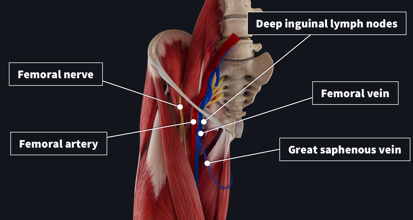
Jul 16, 2021 · Lymph Node Anatomy. Lymph nodes range in size from 1-2 centimeters and are sometimes found alone, or in groups. These bean-shaped structures are composed of four main layers - the capsule ...
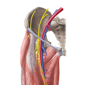
Hip and thigh: Arteries, veins and nerves (preview) - Human ...
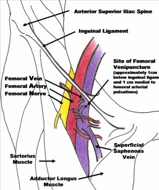
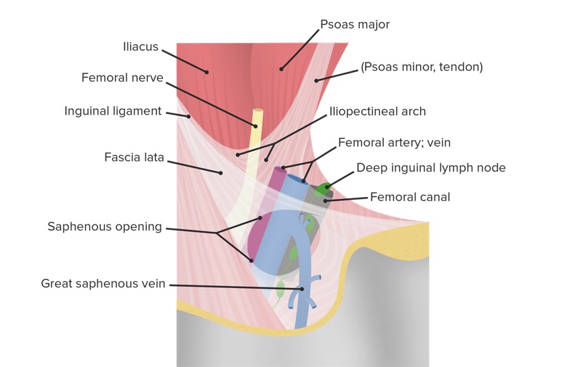
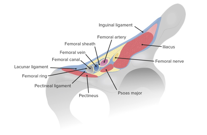
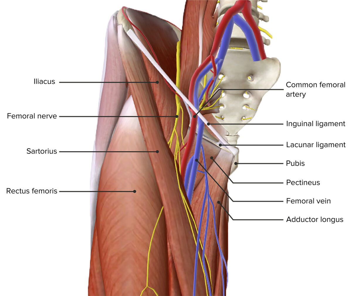
The inguinal lymph node resides within the femoral triangle. The femoral triangle is bounded by the inguinal ligament, adductor longus muscle, and sartorius muscle. The fascia lata forms the roof of the femoral triangle. The floor of the femoral triangle forms from the iliopsoas and pectineus muscles.
Femoral Region and Hernias: Anatomy - Lecturio Medical
Okay, let's know a little about these branches of the dog femoral arteries. The superficial circumflex femoral artery is a small branch of the femoral artery. It arises from the lateral surface of the femoral artery close to the lateral circumflex femoral artery. In a dog, the lateral femoral artery is also known as the cranial femoral artery.
FemoralCanalComplete
The area between the abdomen and the thigh on either side of the body is known as the groin. Anatomical map of center of coordination and centers of fusion of a female pelvic floor identify the follicles corpus albicans and corpus luteum if present lymph nodes groin female diagram.
Cureus | Osteonecrosis of the Femoral Head: Etiology ...
It ends at the popliteal notch of the tibia bone just caudal to the intercondyloid area of the tibia. Again, the femoral ligament of the lateral meniscus is the only femoral attachment of the meniscus in dog knee anatomy. It passes from the caudal axial angle of the lateral meniscus to the medial femoral condyle.

289 Femoral Vein Photos and Premium High Res Pictures - Getty ...
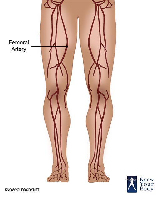
The location of the femoral artery is at the top of your thigh in an area called the femoral triangle. The triangle is just below your groin, which is the crease where your abdomen ends and your legs begin. The femoral artery runs to the lower thigh and ends behind the knee. At the knee, the femoral artery becomes the popliteal artery.
Femoral Artery - Location, Anatomy, Branches, Function and FAQs
The femoral vein is a vein running alongside the femoral artery. The femoral artery is located in the upper area of the thigh and consists of multiple arteries. The deep femoral vein (also known ...

Femoral Artery and its branches - Anatomy tutorial
Download scientific diagram | Arthroscopic view of the intercondylar notch area through the anterolateral portal visualizing the expanded femoral tunnel through the lateral femoral condyle of the ...
Meralgia Paresthetica: Treatment, Diagnosis, Symptoms & Causes
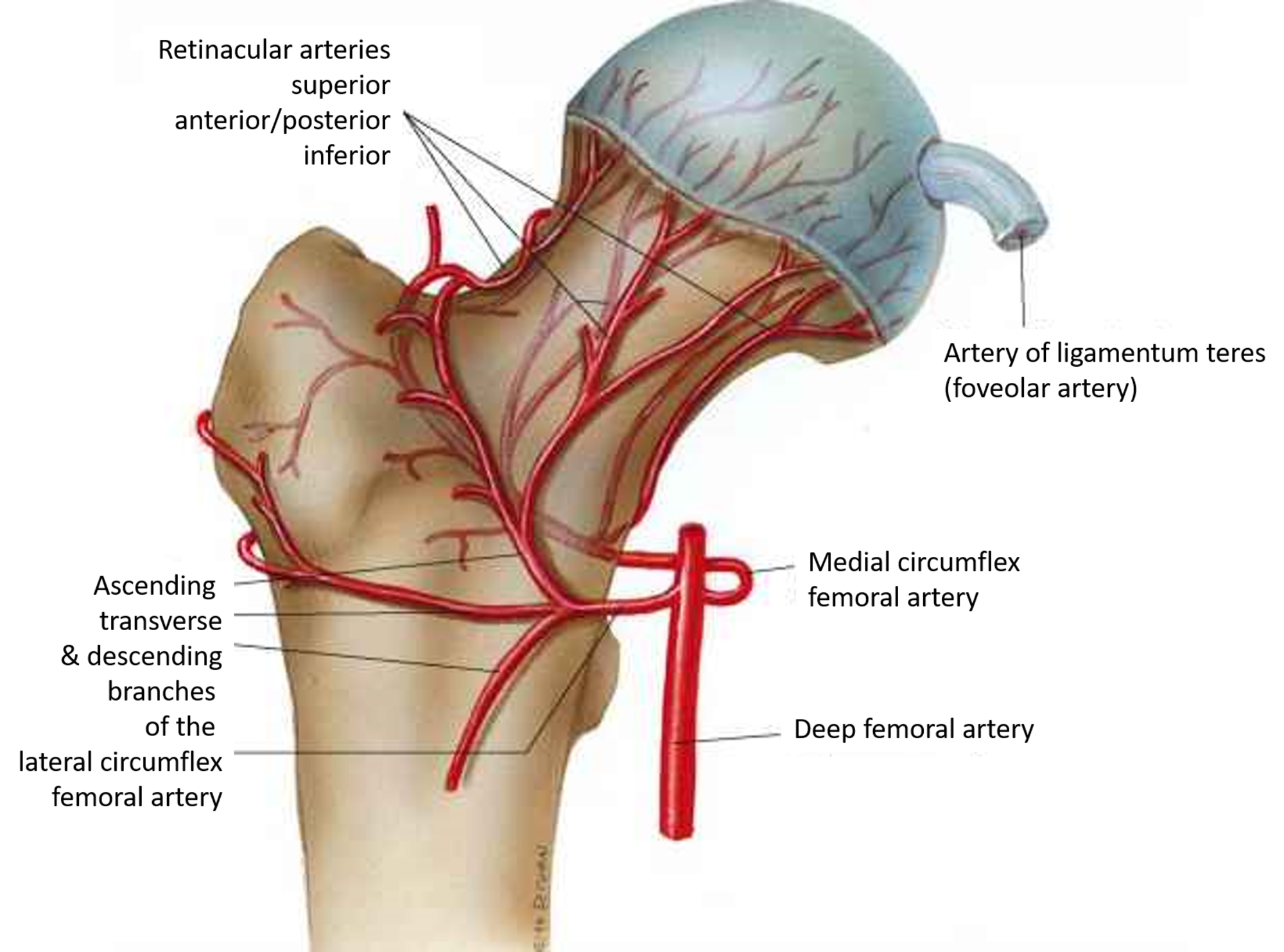
You can feel its pulse in your groin area. It travels from deep within the hip down the thigh and down to the knee. It is the continuation of the external iliac artery which lies within the pelvis. The main blood supply to the femoral head comes from vessels that branch off of the femoral artery: the lateral and medial femoral circumflex arteries.
/GettyImages-87302280-83604c7a3ca84315a84304a002377404.jpg)
Femoral Vein: Anatomy, Function, and Significance
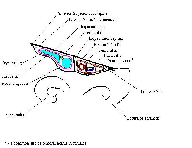
Femoral hernias occur just below the inguinal ligament, when abdominal contents pass into the weak area at the posterior wall of the femoral canal.They can be hard to distinguish from the inguinal type (especially when ascending cephalad) [clarification needed]: however, they generally appear more rounded, and, in contrast to inguinal hernias, there is a strong female preponderance in femoral ...
Femoral triangle: Borders, contents and mnemonics | Kenhub
Aug 21, 2018 · This is a semicircular structure that attaches to two-fifths of the mitral valve’s area. ... Explore the interactive 3-D diagram below to learn more about the mitral valve. ... The femoral ...
The Anatomy of Femoral Vascular Access — Taming the SRU
Meralgia paresthetica is a medical condition resulting from compression (pressure on or squeezing) of the lateral femoral cutaneous nerve (LFCN). This large nerve supplies sensation to the front and side of your thigh. Meralgia paresthetica results in sensations of aching, burning, numbness, or stabbing in the thigh area.

Central line (femoral) - Saving Lives With Sound
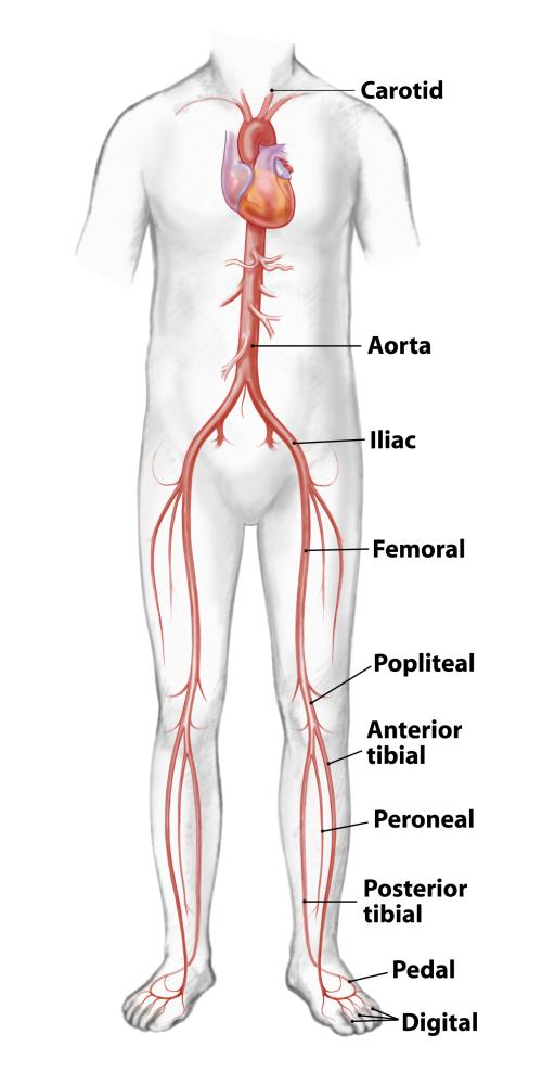
It becomes the femoral artery and branches off as the popliteal artery and the anterior and posterior tibial arteries. The femoral artery supplies blood to the thigh, the popliteal artery supplies the knee area, and the anterior and posterior tibial arteries supply the area below the knee, including the feet and toes.
Femoral Region and Hernias: Anatomy - Lecturio Medical
Sep 02, 2019 · We are pleased to provide you with the picture named Labelled Diagram Of The Muscles In The Human Body.We hope this picture Labelled Diagram Of The Muscles In The Human Body can help you study and research. for more anatomy content please follow us and visit our website: www.anatomynote.com.

Femoral nerve anatomy - UpToDate
The femoral triangle is a wedge-shaped area located within the superomedial aspect of the anterior thigh. It acts as a conduit for structures entering and leaving the anterior thigh. In this article, we shall look at the anatomy of the femoral triangle - its borders, contents, and clinical relevance. Fig 1 - Surface anatomy of the femoral triangle.

Free body diagram around the femoral assembly including the 6 ...
The femoral triangle is formed by the lateral border of adductor longus, the medial border of sartorius and the inguinal ligament (with pectineus and illiopsoas forming the floor). It contains, from lateral to medial, the femoral nerve, artery and vein.
Remember the contents of the Femoral triangle with this ...
Start studying Arteries & Veins. Learn vocabulary, terms, and more with flashcards, games, and other study tools.
Femoral nerve: Anatomy and clinical notes | Kenhub
32) Where on the diagram is the femoral area? a) D b) E c) F d) J e) K Answer: a Difficulty: Medium Learning Objective 1: LO 1.5 Describe the anatomical position and how anatomical terms are used to describe the human body.

Lower Limb Anatomy: The Femoral Triangle - Ponder Med
The femoral head forms the "ball" in the ball and socket joint of the hip. It is also located within the joint capsule and is covered by a synovial membrane. Head of femur Caput femoris 1/3 The femoral neck is about 5 cm long and can be subdivided into three regions.
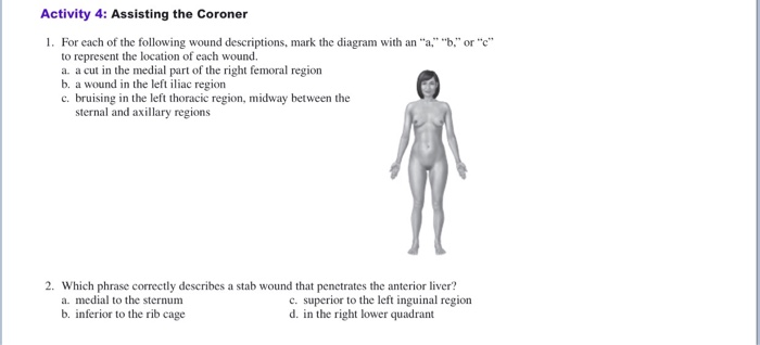
Solved Activity 4: Assisting the Coroner 1. For each of the ...

Femoral Artery - an overview | ScienceDirect Topics
Vascular Anatomy | LeMaitre
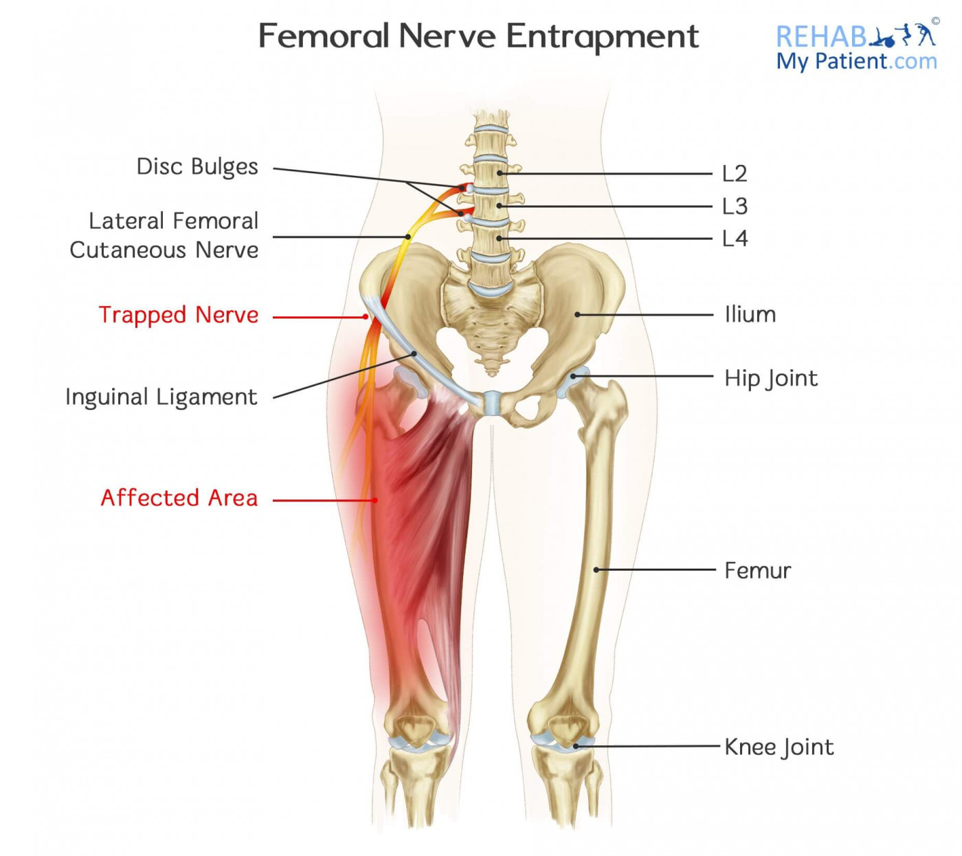
Femoral Nerve Entrapment | Rehab My Patient

Unit 1 Picture questions Flashcards | Quizlet

Hip Pain Explained - including structures & anatomy of the ...
Femoral Head Ostectomy Fho In Dogs | VCA Animal Hospitals
Knee Pain due to Patellofemoral Disorders & Treatments | HSS

Femur Bone Anatomy: Labeled Diagram, Quiz, Color-Coded Parts ...

Femoral artery | Radiology Reference Article | Radiopaedia.org

Figure, Inguinal Region, Inferior Epigastric Artery ...
Femoral artery: Anatomy and branches | Kenhub
Femoral Vein High Resolution Stock Photography and Images - Alamy
0 Response to "37 where on the diagram is the femoral area?"
Post a Comment